

Illustrative consultation about three-dimensional dental imaging
General & Family Dentistry

CBCT 3D Dental Scans

A three-dimensional image is useful when it answers a specific question. Discuss CBCT dental scans in Milton, including why imaging may be recommended, what it can show, and how the findings inform care.

A closer look at CBCT 3D dental scans

Begin with the question the image needs to answer.

Cone-beam computed tomography, usually shortened to CBCT, uses X-rays to create three-dimensional information about teeth and surrounding structures. It can help answer selected questions that an examination and conventional dental images do not adequately resolve. It is not the same as a surface scan used to make a digital impression of the teeth.

The reason for imaging matters more than the appeal of a detailed picture. A clinician may need to understand a particular anatomical relationship or investigate a problem before recommending treatment. The scan should be justified by that need, with an explanation of alternatives, limitations, and radiation exposure.

Contact Milton Dental Clinic to discuss a scan recommendation or the role of imaging in your care. Bring any referral and details of existing images. Ask where the scan would be performed, who will interpret it, and how the report will reach the clinician managing treatment. An online enquiry does not confirm on-site scanning, an imaging appointment, or the suitability of a scan.

Discuss a dental imaging recommendation
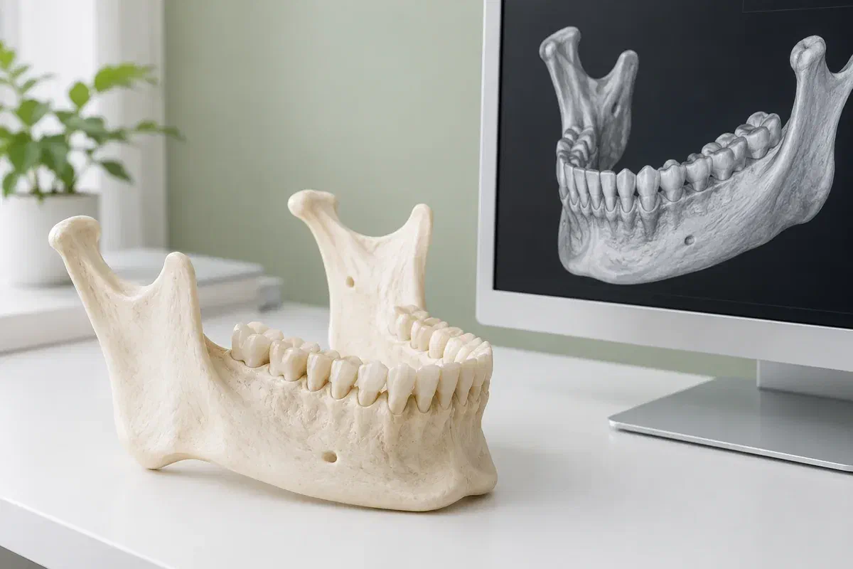
Illustrative jaw teaching model beside a generic three-dimensional dental rendering
Questions that may lead to 3D imaging

Use additional detail where it can change a decision.

CBCT is considered for selected clinical situations. The examples below describe possible reasons for discussion, not a requirement that everyone with these concerns have a scan.

Planning around nearby structures

Some implant or surgical planning requires an understanding of bone and the position of important anatomical structures. Ask which relationship needs clarification and how the information would affect the proposed treatment, including whether another approach remains an option.

Investigating a complex tooth problem

Selected root canal or other dental concerns may warrant further imaging when existing findings leave an important question unresolved. A scan is one part of diagnosis. Symptoms and examination findings still matter, and the image may not provide a definitive explanation.

Clarifying an unusual finding

An unerupted tooth or another finding may need additional assessment of its position or extent. The clinician should explain why the proposed field of imaging is appropriate. Greater coverage is not automatically more useful for a focused dental question.

CBCT should not be treated as a routine upgrade at every check-up. Ask whether an existing image or a lower-radiation examination could provide the information needed.

What the extra dimension can add

See relationships that a flat image may not explain.

Three-dimensional information can help a qualified clinician examine an area from different perspectives. When appropriately selected, it may support diagnosis, planning, or evaluation. That potential benefit needs to be weighed against the exposure and the question being asked.

CBCT uses ionizing radiation and generally involves more exposure than conventional dental X-rays. Dose varies with the equipment, settings, and area examined. The aim is an appropriately limited examination that provides adequate diagnostic information, rather than a promise of zero risk or a fixed dose for every patient.

Patient information: FDA — dental cone-beam computed tomography

A defined planning purpose

Ask what decision depends on the result. A useful explanation connects the image with a clinical choice, such as understanding anatomy before a procedure, rather than simply describing the technology as advanced.

Interpretation within the wider assessment

A scan is reviewed alongside your history, examination, and other records. Ask who is responsible for interpreting the examination and how any findings beyond the original question will be communicated.

Information that can be shared appropriately

A referral may involve an imaging provider and a treating clinician. Clarify how images and the report will be transferred with appropriate authorization so the next appointment can use the completed examination.

From recommendation to results

Know who is arranging each part of the process.

A scan appointment and a treatment consultation may be separate. Confirm the provider, location, fee, and reporting arrangements before travelling, especially if the request came from another practice.

  1. Confirm the indication and existing records

    The responsible clinician reviews why imaging is being considered and whether earlier images answer the question. Provide the dates and locations of previous scans if available. Ask what additional information is expected and how it might change the treatment plan.

  2. Discuss preparation and individual needs

    Follow the imaging provider's instructions. Tell the team if you are pregnant or may be pregnant, have difficulty keeping still, or need positioning assistance. Metal objects near the imaging area may need to be removed as directed.

  3. Acquire the prescribed examination

    The imaging team positions you and explains how to remain still while the equipment moves around the head. The details depend on the device and examination. Raise discomfort or concerns before acquisition so the team can consider whether adjustments are needed.

  4. Review the report with the treating clinician

    The images require interpretation, not just a quick look at a screen. Confirm when results will be available and who will explain them. Further assessment or a change in treatment may be recommended depending on the findings.

After the imaging appointment

Make sure the result reaches the next decision.

The scan itself is an information-gathering step. Its value comes from appropriate interpretation and follow-up, especially when the appointment takes place somewhere other than the practice managing your care.

Confirm the follow-up arrangement

Ask whether you should book a results discussion or wait for the referring clinician to contact you. If a treatment appointment depends on the report, confirm that it has arrived before assuming the plan is ready to proceed.

Keep a record of the examination

Note the date, provider, and area scanned. Tell another clinician about existing imaging before a new examination is proposed. An earlier scan may or may not be sufficient, but reviewing it can help avoid unnecessary duplication.

Report symptoms independently of the scan

A booked or completed scan does not replace urgent assessment of worsening pain, swelling, or another new problem. Tell the treating team if symptoms change while you are awaiting results so the urgency of care can be reconsidered.

Choosing the right information

More detail is helpful only when it is needed.

The choice of imaging should follow the clinical question. Ask whether the expected benefit justifies the proposed examination and whether a different approach could answer the same question.

When existing information is adequate

Examination and available images

History, examination, and current records may be enough to make a decision. A new scan is not automatically necessary because treatment is being discussed. The clinician should explain any uncertainty that remains.

When a two-dimensional view can answer the question

Conventional dental radiography

Selected intraoral or panoramic images may provide the required information. Their role differs from a CBCT examination. Ask why a particular view is proposed and whether it offers sufficient detail for the planned assessment.

When three-dimensional information is necessary

A justified CBCT examination

A qualified provider selects an appropriate examination and reporting pathway. The area and settings should reflect the diagnostic need and patient factors. A scan should not be ordered solely because a larger or more detailed image is available.

Questions before a 3D dental scan

Understand the reason, the exposure, and the follow-up.

Bring the referral or recommendation you received. The discussion can help clarify what is being requested and which clinician or imaging provider will answer practical questions about the examination.

(905) 876-4488
Is a CBCT scan the same as a digital impression for a crown?
No. CBCT uses X-rays to obtain three-dimensional information about internal dental and jaw structures. A digital impression records surface shape using a different type of scanner. Ask which examination has been recommended, because the purpose, preparation, and exposure considerations are different.
Does every implant consultation require a new CBCT appointment immediately?
The imaging plan follows assessment of the proposed treatment and available records. A clinician may need three-dimensional information, but the timing and whether a new scan is required should be explained. Bring earlier images or their details rather than arranging duplicate imaging independently.
Can a dental CBCT scan be described as radiation-free?
No. It uses ionizing radiation. The responsible clinician should explain the expected benefit, alternatives, and how the examination is limited to the information needed. Dose varies, so a single advertised comparison does not describe every scan or every patient.
What should I say if I might be pregnant before dental imaging?
Tell the referring clinician and imaging team before the examination. They can consider the clinical need, timing, and relevant precautions for your situation. Do not assume that a necessary assessment should be cancelled without discussing the reason for the imaging.
Who explains findings on a scan performed at another imaging centre?
Clarify this before the appointment. The scan requires appropriate interpretation and a report that reaches the clinician managing your care. Ask who will contact you, when to expect a discussion, and how additional findings or recommendations will be followed up.
Can I book a scan through the website without a clinical assessment?
Use the enquiry to discuss the recommendation and any existing referral. A website request does not establish that CBCT is indicated or confirm where it will be performed. The team can clarify the appropriate assessment or referral pathway and the arrangements that need confirmation.

Patient information: RCDSO — dental CT scanner requirements

CBCT 3D dental scans in Milton

Let’s clarify what the recommended image is for.

Contact our Bronte Street practice with your imaging questions and any referral details. We can discuss the role of a scan in your dental assessment and clarify the next step. Confirm the imaging provider and appointment arrangements before attending for a scan.

Online requests are enquiries, not confirmed scan bookings. Call if a painful or swollen area needs prompt assessment.

Visit Milton Dental Clinic
Unit 1, 342 Bronte St. S, Milton, ON L9T 5B7
Get directions Clinic hours
Monday–Thursday
11am–7pm
Friday–Sunday
By appointment only

How can we help?

Milton Dental Clinic

Have a question about your smile? Choose the option that works best for you.

For urgent dental concerns, please call.
Messages are reviewed during clinic hours.