

Illustrative consultation about three-dimensional dental imaging
General & Family Dentistry

CBCT 3D Dental Scans in Georgetown

A tooth that still aches after treatment, with nothing obvious on the usual X-ray, is frustrating. From Georgetown, find out how a CBCT 3D dental scan can add information to that kind of puzzle, where its limits lie, and what comes next.

An unexplained tooth, Georgetown

When the usual X-ray comes back quiet

You know which tooth is bothering you. It may have had a root canal or a large filling, and it still feels tender when you bite. Yet the X-ray looks unremarkable. In some cases like this, a dentist or specialist may consider cone-beam computed tomography, CBCT, to look at the tooth and surrounding bone in three dimensions.

CBCT is an X-ray exam, not the optical camera used to make a digital impression. It can add depth that a flat image lacks, but it does not explain every symptom. A scan should be proposed for a specific diagnostic question, with an honest account of the alternatives, the limits of imaging and the radiation it involves. Your description of the pain and the clinical tests remain just as important.

Contact Milton Dental Clinic to discuss an imaging recommendation for a troublesome tooth. Bring any referral and a note of earlier X-rays or scans. Ask where the scan would be taken, which qualified person reads it, and by what route the findings get to whoever is treating you. An online enquiry does not confirm imaging, on-site scanning or suitability.

Discuss a scan for a problem tooth
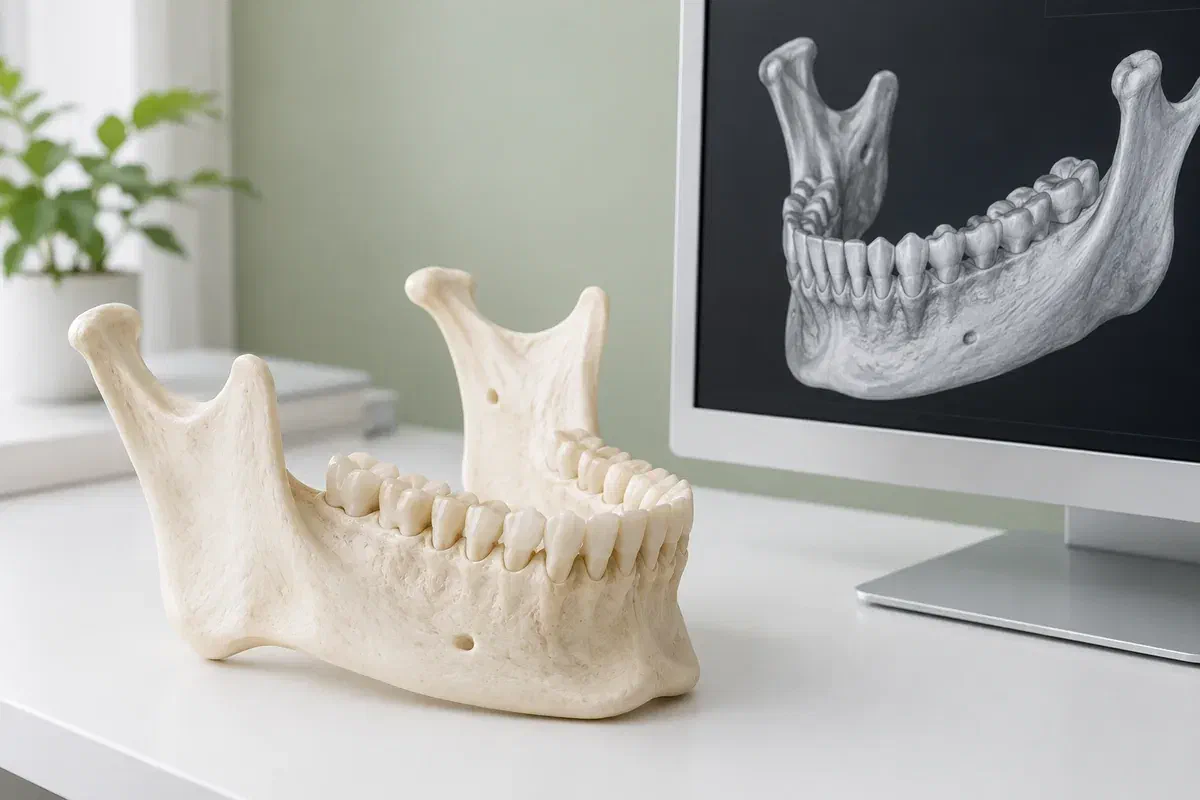
Illustrative jaw teaching model beside a generic three-dimensional dental rendering
When a clinician may suggest CBCT

Reasons to look deeper

A 3D scan is sometimes raised for the situations below. Each is a reason to discuss imaging, not a sign that a scan is certain to help.

Anatomy before surgery or an implant

If a problem tooth may eventually be replaced, or surgery is being considered, understanding bone and nearby nerves or sinuses can matter. Ask what is unclear, how the result would affect the plan, and whether other treatment would remain possible.

Ongoing symptoms after root canal treatment

When pain or tenderness persists and routine images do not explain it, a clinician may want more detail about the roots and surrounding bone. The scan is one input among several, and it may not reveal a definite cause.

Something unclear on a previous image

A shadow near a root, or a tooth that never came through, can leave its edges and depth uncertain. Ask why the proposed area of imaging is suitable. A larger field does not necessarily help with a focused question.

CBCT is not a routine next step for every sore tooth. Ask whether current X-rays, or a different conventional view, could provide the information needed.

What depth may reveal

Another angle on a stubborn problem

Flat X-rays overlap structures, which can hide detail. CBCT lets a qualified clinician view the tooth and bone in cross-sections from several directions. When chosen for a clear reason, that may support diagnosis, treatment planning or evaluating how an area has responded. It still has to justify its exposure.

CBCT uses ionizing radiation and usually involves a higher dose than routine dental X-rays. The amount depends on the equipment, its settings and the size of the region scanned. Good practice keeps the scan limited to the area in question while still producing diagnostically adequate images. That is not the same as being risk-free, and there is no single dose that applies to every scan.

Patient information: FDA — dental cone-beam computed tomography

A decision tied to the images

Ask what will be decided differently once the scan is reviewed, for instance whether to retreat a tooth, refer you or consider other options. A good explanation links the image to that choice.

Images read alongside your symptoms

Interpretation considers your history, clinical tests and other records. Ask who will read the scan and how they will tell you about anything they notice beyond the original question.

Findings that reach your treating clinician

If a separate provider takes the scan, check how the files will be passed along with your authorization, so your next appointment can use the completed exam.

What the process looks like

Four stages, one question

Imaging and any treatment talk may land on different days, even at different addresses. Before leaving Georgetown, know who is scanning you, where, what it costs and who receives the report.

  1. Review what has been tried

    Your clinician weighs the reason for 3D imaging against what earlier films already show. Tell them about any previous scans, including the office that took them. Ask what the new images are expected to add.

  2. Get ready

    Follow the imaging provider’s instructions. Mention a possible pregnancy, difficulty holding still or any need for positioning help. You may be asked to take off jewellery, glasses or removable dental appliances.

  3. Have the images taken

    You will be positioned and asked to stay still while the unit moves around your head. Exact steps depend on the machine and the area. Raise discomfort or worries before the scan starts.

  4. Hear the interpretation

    The images need a qualified reading, not a quick glance. Check the expected timing and the name of the person who will sit down with you over the findings. Further tests, a referral or a revised plan may follow.

While you wait, and after

Keep the pain and the report connected.

A scan collects information without treating the tooth. Its value depends on interpretation and follow-up, which needs a little more coordination when imaging happens away from the office managing your care.

Know how you will hear back

Ask whether to book a results visit or wait for the referring clinician to contact you. When retreatment or another procedure is waiting on the findings, confirm your dentist has them in hand first.

Track the symptom and the scan

Keep a simple record of when the tooth hurts, along with the date, provider and area of the scan. If another clinician suggests imaging later, mention this exam so it can be reviewed first.

Don’t wait out a worsening tooth

A booked or completed scan is no reason to delay care if pain increases, swelling develops or something new appears. Tell the treating team about changes so they can reconsider how soon to see you.

Imaging options for a problem tooth

Not every mystery needs a 3D scan.

The clinical question should guide the imaging. Ask whether the expected benefit justifies a CBCT and whether a simpler method could answer the same question.

When the existing record is enough

Clinical tests and current images

Your history, the exam, tests such as tapping or cold, and recent X-rays may be sufficient to decide. A treatment discussion does not automatically mean a new scan. The clinician should explain what is still uncertain.

When a two-dimensional image can help

A conventional view from another angle

An additional periapical image taken at a different angle sometimes reveals what the first one missed. Ask what the proposed view is meant to show and whether it gives enough detail.

When three dimensions are needed

A small-field CBCT

For a single tooth, a limited field focused on that area is often what a qualified provider would choose, with a proper report to follow. Settings should suit the question and you. A scan should not be ordered just because a larger or more detailed image is possible.

Questions about a problem tooth

What a scan can and cannot tell you

Bring your referral and any notes about previous treatment on the tooth. A short conversation usually shows what is being asked for, and which practical details belong with the imaging office instead.

(905) 876-4488
Can a CBCT find a crack in a tooth?
Sometimes it shows signs associated with a crack, but fine cracks are often not visible on imaging. Diagnosis usually relies on symptoms, clinical tests and examination as well. Ask your dentist what they hope the scan will clarify.
Will the scan tell me whether to retreat the root canal or remove the tooth?
It may provide information that helps with that decision, but the choice also depends on your symptoms, the tooth’s condition, your preferences and the options available. Ask how the findings would affect each choice.
Do metal posts or fillings interfere with the images?
Metal and some dental materials can cause streaks or shadows that limit what is visible nearby. That trade-off is part of judging whether 3D imaging is worthwhile for your particular tooth.
Should I keep taking pain medication before the scan?
Follow the advice of your dentist, physician or pharmacist about medication. The scan itself does not usually require changes. If pain is getting worse, contact the treating team rather than waiting for the appointment.
Does the scan replace the tests my dentist already did?
No. Imaging works alongside clinical tests and your history. A scan may add detail, but it cannot replace an examination or explain symptoms on its own.
Is a referral needed for a dental CBCT?
A scan should follow a clinical assessment that identifies a reason for it. Use an enquiry to share your referral or recommendation, and the team can explain the appropriate pathway and what needs to be arranged.

Patient information: RCDSO — dental CT scanner requirements

Georgetown tooth-pain imaging questions

Bring the history of that tooth.

Tell our Bronte Street team what has already been done on the tooth, and share any referral you were given. Together we can look at whether a scan belongs in the assessment and what should happen after it. Confirm the imaging provider and appointment before attending.

Online requests are enquiries, not booked scans. Call if a tooth is becoming more painful or swelling appears.

Visit Milton Dental Clinic
Unit 1, 342 Bronte St. S, Milton, ON L9T 5B7
Get directions Clinic hours
Monday–Thursday
11am–7pm
Friday–Sunday
By appointment only

Areas we serve

  • Milton
  • Oakville
  • Mississauga
  • Georgetown
  • Halton Hills
  • Acton
  • Campbellville
  • Brampton
  • Burlington
  • Puslinch
  • Rockwood
  • Erin
  • Caledon
  • Etobicoke
  • Old Milton
  • Bronte Meadows
  • Timberlea
  • Dorset Park
  • Dempsey
  • Clarke
  • Beaty
  • Coates
  • Harrison
  • Scott
  • Willmott
  • Ford
  • Bowes
  • Cobden
  • Walker
  • Mountain View
  • Milton Heights
  • Hawthorne Village
  • Valley View
  • Fallingbrook
  • Nassagaweya
  • Trafalgar
  • Esquesing
  • Nelson
  • Brookville
  • Moffat
  • Glen Williams
  • Norval
  • Limehouse
  • Stewarttown
  • Ashgrove
  • Hornby
  • Silver Creek
  • Speyside
  • Scotch Block
  • North Oakville
  • River Oaks
  • Glen Abbey
  • West Oak Trails
  • Palermo
  • Bronte Creek
  • College Park
  • Joshua Creek
  • Uptown Core
  • Preserve
  • Iroquois Ridge
  • Lisgar
  • Meadowvale
  • Meadowvale Village
  • Churchill Meadows
  • Erin Mills
  • Central Erin Mills
  • Streetsville
  • East Credit
  • Creditview
  • Heartland
  • Sheridan
  • Mount Pleasant
  • Credit Valley
  • Huttonville
  • Churchville
  • Springbrook
  • Northwest Brampton
  • Bram West
  • Fletcher's Meadow
  • Fletcher's Creek South
  • Alton Village
  • Orchard
  • Millcroft
  • Headon Forest
  • Tansley
  • Brant Hills
  • Tyandaga
  • Kilbride
  • Lowville
  • Aberfoyle
  • Morriston
  • Arkell
  • Hillsburgh
  • Freelton
  • Carlisle
  • Waterdown
  • Flamborough
  • Mountsberg

How can we help?

Milton Dental Clinic

Have a question about your smile? Choose the option that works best for you.

For urgent dental concerns, please call.
Messages are reviewed during clinic hours.