A tooth edge your tongue keeps finding
Close-ups can show a chip or a worn area that feels sharp. They do not show how deep the change is or explain its cause. Mention any sensitivity, grinding or bite changes so the finding is understood in full.


That one spot that bleeds every time you floss is usually somewhere you cannot see. A magnified photo from an intraoral camera can show where it is, so the cleaning advice you take home is aimed at the right tooth and the right edge of the gum.
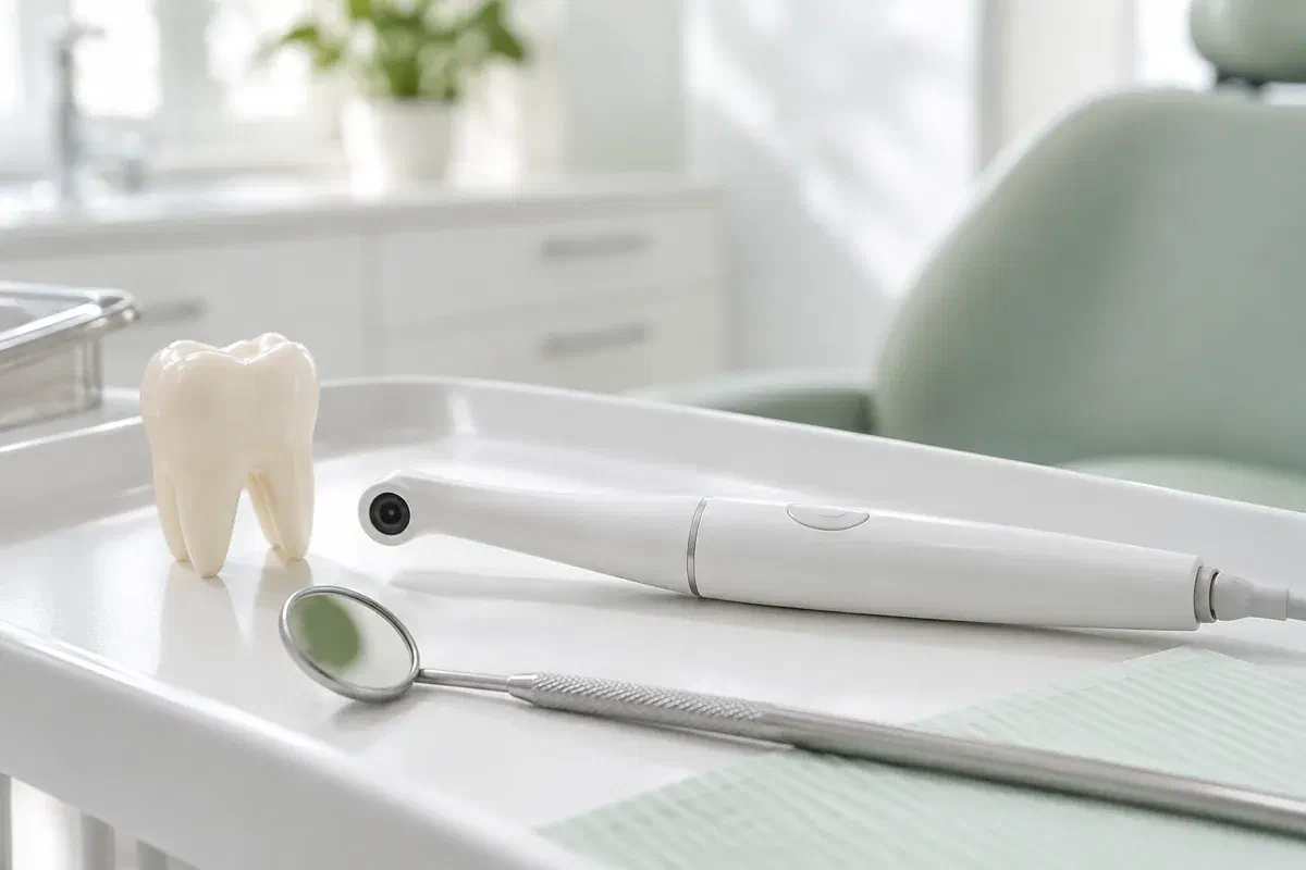
Picture the inside of your last molar, the side that faces your tongue. Most of us never have. When a dentist says plaque is building up there, or that a filling edge looks rough, it helps to have something better than imagination. An intraoral camera is a slender handheld camera that takes enlarged photos of reachable teeth and gum surfaces and displays them on a screen.
Those pictures help the conversation; they do not make the diagnosis. Glare, moisture, the angle of the lens and how much of the area is exposed all change the result. Your dentist has to put the image together with your symptoms, your history and the rest of the examination before explaining its meaning or suggesting treatment.
Planning a visit from Burlington, you are welcome to ask whether photos would help with the concern you are bringing and what arrangements are available. In the chair, ask which tooth is on screen, what feature matters and what the image cannot tell. A close-up is meant to make the next step clearer, not to alarm you because a tooth looks unfamiliar when magnified.
A photo is most helpful when it is tied to a clear question. Mention the area that concerns you, and ask how the image relates to everything else the dentist finds.
Close-ups can show a chip or a worn area that feels sharp. They do not show how deep the change is or explain its cause. Mention any sensitivity, grinding or bite changes so the finding is understood in full.
A photo may reveal a ledge or gap where food tends to collect. A visible line still has to be assessed in person. Ask what further checks are needed before deciding the filling should be replaced.
Repeated bleeding at one site often points to a spot where cleaning is falling short. A photo can show that location clearly. Ask the team to connect it with a demonstration using your own brush or floss, because seeing the spot only matters if you can reach it afterward.
Close-up photographs differ from dental X-rays and from three-dimensional digital impressions. Each kind of record has its own job, and they cannot be used in place of one another.
Your dentist can point to the precise surface on screen while explaining why it needs attention. For gumline areas, back teeth and tight contacts, that can turn vague advice into something specific. Do not feel you need to analyse every highlight or colour; ask which section of the picture to focus on.
Images may be saved to your record where appropriate. Putting an older photo beside a newer one can help a review, but different angles and lighting can make one tooth look like two. Comparisons only make sense together with the exam and your history, not as proof of change by themselves.
Patient information: Carestream Dental — intraoral camera image capture and review
Ask which tooth and which surface are shown. Knowing the location makes it easier to clean the area properly and to describe any symptoms later. An enlarged image may take a moment to orient, so ask which way is up and how large the area really is.
If treatment is suggested, the photo lets you ask what it would fix and which findings support it. Other options may exist. The picture is one part of the explanation and should not shorten the discussion of benefits, limitations and choices.
If the plan is to watch an area, ask what will be compared at your next visit. Photographs may help together with gum measurements, symptoms and other records. Ask, too, which changes should prompt an earlier call.
The camera is a small supporting step inside a regular clinical visit. Let the team know about tender gums, a sensitive gag reflex or a jaw that does not open far, so pictures can be taken comfortably.
You explain what brought you in and the dentist examines the area. Ask what a picture could add. Some problems cannot be judged by appearance, and photographs never replace a proper assessment.
The slim camera is angled toward the tooth or gum surface. The team might reposition it or dry the area for a sharper image. Signal if you need a pause or the position is uncomfortable.
Ask the dentist to indicate the relevant feature and to separate what is seen from what is concluded. A photo may show where something is without telling you its depth, whether it is active or why it began. Other findings complete the explanation.
That could be home-care advice, a plan to monitor, more tests or a conversation about treatment options. Confirm the reason and the timing of any follow-up. If images are kept or shared for your care, ask how they are recorded and what consent applies.
Having photographs taken does not normally need recovery care. What counts afterward is the advice or plan drawn from the full exam.
If the image revealed a spot you have been missing, use the method demonstrated for that area. Ask for another demonstration at your next visit if it still feels awkward. Remember that a picture cannot clean a tooth for you.
Before leaving, confirm what was seen, what it suggests and what is still uncertain. Ask about terms you did not follow. A magnified gumline can look much worse than it is, so the explanation is what matters.
If the area is being monitored, note the next check and which symptoms mean calling earlier. A saved image does not replace future exams. New pain, swelling or a change in how a tooth works deserves attention even if a past photo seemed reassuring.
You are not expected to read dental images. The dentist should explain how the picture fits the exam, be honest about what is uncertain and answer your questions about what comes next.
Contact our Bronte Street practice and tell us which area has been bothering you. We can let you know whether close-up photos are likely to help and how bookings work. We will explain what the images show, what they cannot establish and how to look after the area at home.
Photographs support a clinical assessment. The illustrative images on this page do not show the clinic’s equipment.
