

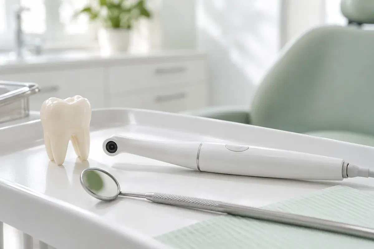
Illustrative dental consultation with a small intraoral camera, not actual clinic equipment
General & Family Dentistry

Intraoral Camera Examinations in Puslinch

A brown fleck in a back groove, a grey shadow near a filling: dark marks on teeth worry people, and many turn out to be harmless. An intraoral camera can enlarge the spot on screen so your dentist can explain what it most likely is and what the rest of the exam shows.

Puslinch · a closer look at dark spots

Stain, shadow or something more?

It usually starts with a glance in the mirror or a photo on someone’s phone: a darker patch on a molar you had not noticed before. Coffee, tea, natural pits in the enamel, the rim of an old filling and early decay can all look similar from a distance. An intraoral camera is a small handheld lens with its own light that captures a magnified view of reachable tooth surfaces, so the mark can be examined together.

Bear in mind that colour on a screen is not a diagnosis. Glare, saliva, the angle of the shot and the camera’s lighting all influence how dark something looks. Your dentist puts the photo beside your symptoms, history and the hands-on exam, and may suggest other checks, before saying what the spot is.

For households in Puslinch, west of Milton, it often makes sense to raise the concern when you call and ask whether photographs would help and how the visit can be set up. At the appointment, ask which tooth is shown, what the mark appears to be and how confident that impression is. A large, dark image on a screen should lead to an explanation, not a snap decision.

Have a dark spot checked
Illustrative pen-shaped intraoral camera beside a tooth model and dental mirror
Common sources of worry

Marks that often prompt a closer look

Photos are most helpful with a specific question in mind. Point to the mark that concerns you and ask how the image compares with other findings.

Darker grooves or a worn surface

Natural fissures can pick up stain, and worn enamel can look darker than its surroundings. A close-up may show the pattern, but not how deep any change goes. Mention sensitivity or food catching so the dentist can judge the spot properly.

A shadow beside an older filling

Restoration edges sometimes appear grey or dark, especially with older materials. That alone does not mean the filling has failed. Ask what further examination would show whether anything is happening beneath the surface.

Discolouration along the gumline

Tartar and stain often collect where brushing is difficult. An image can show the location, and a quick demonstration can help you reach it. The photo is only half the answer; the technique is the other half.

An intraoral photograph cannot do the work of an X-ray or a 3D scan. When a dark spot needs assessing below the surface, a different record may be needed.

What the close-up adds

Context for a colour that caught your eye

With the spot magnified, your dentist can show where it begins and ends and explain which features suggest stain, a natural groove or something that needs attention. Rather than reacting to how dark a pixel looks, ask what in the image actually matters.

If the mark is being monitored, the photograph may be saved to your record where appropriate. Lighting and angle can make the same spot look lighter or darker from one image to the next, so any comparison belongs with a fresh exam and your history, not as stand-alone proof.

Patient information: Carestream Dental — intraoral camera image capture and review

Locating the mark exactly

Find out which tooth and which surface the spot is on. That helps you watch for symptoms and keep the area clean. On a big screen, orientation can be confusing, so ask how the image relates to your mouth.

Understanding what is recommended

If a filling or other care is suggested, the image gives you something concrete to ask about: what would treatment fix, and what evidence supports it? Monitoring may be an alternative for some spots. Talk through benefits and limits.

A baseline for comparison

Some spots are simply kept under observation. In that case, find out what the dentist will compare at your next checkup and what kind of change would prompt action rather than more watching. Photos can help along with other records and any symptoms you report.

During the exam

Looking at the mark step by step

The camera is one tool within a clinical visit. Let the team know about tender spots, a tendency to gag or difficulty opening so photos can be taken comfortably.

  1. Point out what you noticed

    Describe where you saw the mark and how long it has been there. The dentist examines the tooth and explains whether a photo would help. Colour alone cannot answer every question, and the camera does not replace assessment.

  2. Enlarge the spot

    The lens is positioned close to the tooth. A clearer image may need a dry surface or a slight change in angle. Signal if you would like a pause.

  3. Interpret the colour carefully

    Ask which features point toward stain, a groove, a filling edge or decay, and which parts remain uncertain. A photo shows where something is visible, not how deep it runs or whether it is active. Other checks may follow.

  4. Agree on what comes next

    The plan could be reassurance, cleaning advice, monitoring, more tests or treatment. Ask for the reasons and any timing. You can also ask how images are stored and shared with consent if needed.

After the appointment

A dark spot explained is a lot less worrying

Photographs need no recovery care. The value lies in the explanation and plan from the full examination.

Be clear about the verdict

Before leaving, confirm whether the mark is likely stain, a natural feature, a filling edge or decay, and what is still uncertain. Ask about any terms you did not follow. Magnification can make a small spot look large.

Follow advice that fits the cause

Stain may call for cleaning changes, decay may call for treatment and a stable spot may only need watching. Use the guidance for your situation and ask for a demonstration if helpful. The photo itself changes nothing.

Keep the review if one is planned

If the spot is being monitored, note the date and which symptoms mean calling earlier, such as new sensitivity, food catching or pain on biting. A saved image does not replace the next exam, and a spot that looked harmless in one photo still deserves a fresh look if it starts to bother you.

Telling stain from decay

Tools that look at a spot differently

A dark mark can be looked at in more than one way, and your dentist picks records to suit the question being asked. If more than a photo is proposed, it is reasonable to ask what extra information it would give and whether the result could change the advice.

Colour, outline and location

Intraoral photos

A photograph documents what is visible and where. It depends on image quality and interpretation, and colour can shift with lighting. Useful for explanation, not a complete diagnosis.

Whether anything lies beneath

Tests and X-rays where needed

To judge whether a dark area involves decay below the surface, the dentist may recommend clinical tests or radiographs. Ask what they would reveal. A clear photo does not cancel an investigation aimed at a separate question.

If a restoration is planned

Digital impressions

Should a crown or appliance be needed, an optical scanner can capture a 3D model for that work. That is quite different from photographing a spot. Ask which records your treatment would involve.

Dark spot questions

What a photo can and cannot settle

You are not expected to diagnose a spot from a picture. The dentist should explain the evidence, the uncertainty and the reasons behind any recommendation.

(905) 876-4488
Can coffee or tea stains look like decay in a close-up?
Yes. Surface stain can settle into grooves and look very dark when magnified. The dentist considers texture, location and other findings before deciding whether it is stain or decay.
Why would a dentist want an X-ray of a spot they have already photographed?
A photograph shows only the outer surface. A radiograph may help show whether decay extends between teeth or beneath the enamel. Ask what the X-ray is meant to answer.
Can a dark spot be cleaned away during the visit?
Some stains and deposits can be removed during a cleaning, while other marks are part of the tooth or a restoration. The dentist can tell you which applies and what to expect.
Does a small dark spot always need a filling?
No. Some spots are stain or stable features and only need monitoring. The decision depends on the full exam and your risk factors. Ask why treatment or monitoring is being suggested.
Will the photo show if the spot has spread since my last visit?
It may help, but lighting and angle changes can alter appearance. The dentist should combine any comparison with a fresh examination before saying a spot has changed.
Is it worth booking just to ask about one dark mark?
If it concerns you, yes. Mention it when you call. If you are due for a checkup, it can often be looked at then. New pain or sensitivity with the spot is a reason to call sooner.
Booking from Puslinch

Let’s find out what that spot really is.

Phone our Bronte Street practice and describe the mark you have noticed, where it is and roughly when it appeared. The team can tell you whether a camera view is likely to be useful and how to set up the visit around your other plans. Your dentist will go over what the image suggests, what remains uncertain and what, if anything, needs doing.

Camera images support a clinical assessment. Photographs on this page are illustrations and are not of clinic equipment.

Visit Milton Dental Clinic
Unit 1, 342 Bronte St. S, Milton, ON L9T 5B7
Get directions Clinic hours
Monday–Thursday
11am–7pm
Friday–Sunday
By appointment only

Areas we serve

  • Milton
  • Oakville
  • Mississauga
  • Georgetown
  • Halton Hills
  • Acton
  • Campbellville
  • Brampton
  • Burlington
  • Puslinch
  • Rockwood
  • Erin
  • Caledon
  • Etobicoke
  • Old Milton
  • Bronte Meadows
  • Timberlea
  • Dorset Park
  • Dempsey
  • Clarke
  • Beaty
  • Coates
  • Harrison
  • Scott
  • Willmott
  • Ford
  • Bowes
  • Cobden
  • Walker
  • Mountain View
  • Milton Heights
  • Hawthorne Village
  • Valley View
  • Fallingbrook
  • Nassagaweya
  • Trafalgar
  • Esquesing
  • Nelson
  • Brookville
  • Moffat
  • Glen Williams
  • Norval
  • Limehouse
  • Stewarttown
  • Ashgrove
  • Hornby
  • Silver Creek
  • Speyside
  • Scotch Block
  • North Oakville
  • River Oaks
  • Glen Abbey
  • West Oak Trails
  • Palermo
  • Bronte Creek
  • College Park
  • Joshua Creek
  • Uptown Core
  • Preserve
  • Iroquois Ridge
  • Lisgar
  • Meadowvale
  • Meadowvale Village
  • Churchill Meadows
  • Erin Mills
  • Central Erin Mills
  • Streetsville
  • East Credit
  • Creditview
  • Heartland
  • Sheridan
  • Mount Pleasant
  • Credit Valley
  • Huttonville
  • Churchville
  • Springbrook
  • Northwest Brampton
  • Bram West
  • Fletcher's Meadow
  • Fletcher's Creek South
  • Alton Village
  • Orchard
  • Millcroft
  • Headon Forest
  • Tansley
  • Brant Hills
  • Tyandaga
  • Kilbride
  • Lowville
  • Aberfoyle
  • Morriston
  • Arkell
  • Hillsburgh
  • Freelton
  • Carlisle
  • Waterdown
  • Flamborough
  • Mountsberg

How can we help?

Milton Dental Clinic

Have a question about your smile? Choose the option that works best for you.

For urgent dental concerns, please call.
Messages are reviewed during clinic hours.