A chipped or worn contact point
When the edge where two teeth meet wears or chips, a small gap can form. A close-up may reveal the change, though not its depth or cause. Mention sensitivity, grinding or any recent bite change.


Floss that shreds in the same gap, or food that packs in after every meal, usually has a reason you cannot see. A magnified photo from an intraoral camera can show the ledge, gap or worn contact involved while your dentist works out what is causing it.
A strand of meat lodged between the same two molars, a toothpick reached for after every supper, floss that catches and frays: these small irritations are some of the most common reasons people ask for a closer look. The cause might be a rough filling margin, a small space that has opened between teeth or a worn contact. An intraoral camera, a narrow handheld instrument with a light at the end, photographs reachable surfaces up close so the spot can be shown on screen.
Even so, the photograph is a helper. It reflects the lighting, the angle, how wet the area is and how much of the contact the lens can actually reach, which between teeth is often not much. Your dentist combines it with your description, your history, a hands-on exam and possibly other records before explaining what is going on.
From Rockwood, northwest of Milton, it can be useful to describe the food trap when you book and ask whether photographs might help and how the appointment can be organised. Once the image is up, find out which pair of teeth you are looking at, which edge or gap matters and what stays out of view. A dramatic close-up of a filling margin should lead to an explanation, not a rushed choice.
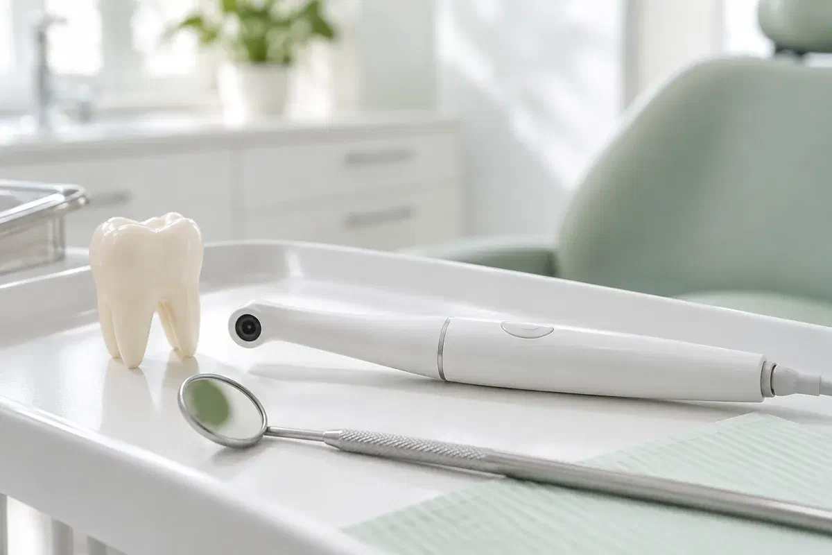
A photo works best when it is aimed at a specific question. Show the dentist where food collects and ask how the image ties in with the rest of the findings.
When the edge where two teeth meet wears or chips, a small gap can form. A close-up may reveal the change, though not its depth or cause. Mention sensitivity, grinding or any recent bite change.
An overhanging or uneven edge can snag food and floss. A photo can show the visible part, but what is happening under or between teeth may need other checks. Ask what those are before replacement comes up.
When gum tissue shrinks back, a triangular space can open where food collects. A photo can show it, and the team can recommend a cleaning aid suited to that space and show you how to use it.
Between-tooth problems are often partly hidden from a camera. Photographs, X-rays and 3D digital impressions each answer different questions and are not interchangeable.
On screen, your dentist can trace the ledge, gap or worn contact that keeps catching food and explain whether it is a concern. That is much easier than describing a spot behind your molars. Rather than inspecting every reflection, ask which part of the picture shows the trap.
Where appropriate, the image can be stored in your record. If the area is being monitored, an earlier photo might be compared with a newer one, but angle and lighting matter. A comparison needs a fresh exam and your history beside it.
Patient information: Carestream Dental — intraoral camera image capture and review
Find out exactly which teeth and which side are involved. That helps you aim floss or a small brush correctly and describe symptoms if they change. Magnified images can be hard to orient, so ask which way you are looking.
Smoothing a margin, repairing a filling or simply changing your cleaning routine may each be possible. The photo helps you ask what a proposed step would solve and which findings support it. Ask about the alternatives too.
If the plan is to monitor the area, find out what will be compared next time and which changes, such as pain or swelling, mean calling sooner. Photos can complement notes and other records.
Photographs are a short part of a clinical exam. If the area is sore from trapped food, or you gag easily or cannot open wide, tell the team so they can work comfortably.
Describe which foods catch, how often and whether the area is tender. The dentist examines the contact and may check it with floss. Ask whether a photo would help; between-tooth concerns are not always visible.
The lens is brought as close as possible to the gap or filling edge. A change in angle or a quick dry may be needed. Signal if you want a break.
Ask what the image actually shows and what it leaves out. A photo can reveal a visible ledge or space without proving what is happening beneath it. Clinical checks or X-rays may add what the camera misses.
Outcomes range from cleaning advice to smoothing, repair, monitoring or further tests. Ask why one is preferred and when to return. You may also ask how photos are kept and whether they would be shared, with consent, for your care.
Having photographs taken does not call for recovery care. What matters next is the advice that comes from the full exam.
Before you leave, check whether it is a filling edge, a space, worn teeth or gum recession, and what is still uncertain. Ask about any unfamiliar words. A magnified ledge can look worse than it feels.
Floss, a small interdental brush or another aid may be suggested for that particular gap. Practise the technique you were taught. A photo does not remove trapped food or replace any repair that was planned.
Keep any review appointment. If the area becomes painful, the gum swells or food packing gets worse, call before the scheduled date. A saved image does not replace a new exam.
You do not have to work out the cause from a photo. The dentist should explain the findings, the uncertainty and the reasoning behind any suggested fix.
Get in touch with our Bronte Street practice and describe where food keeps catching. The team can tell you if camera photos are likely to help and how the visit can be planned. Your dentist will explain what the image shows, what remains hidden between the teeth and what can be done.
Photographs support a clinical assessment. Images on this page are illustrative and do not depict clinic equipment.
