

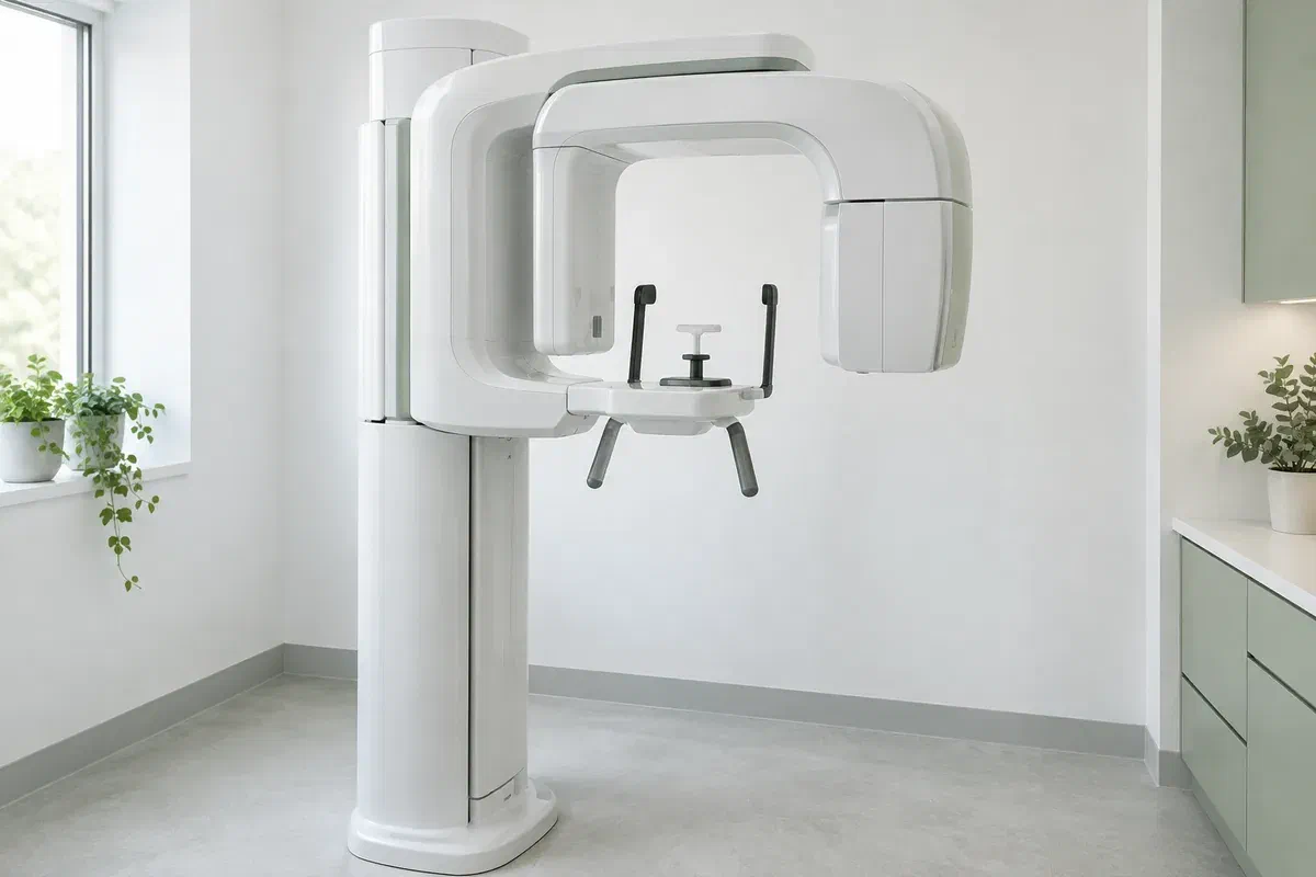
Illustrative discussion near generic panoramic imaging equipment, not actual clinic facilities
General & Family Dentistry

Panoramic Dental X-rays

Understand why a broader view may help your dental assessment. Discuss panoramic X-rays in Milton, the question the image is meant to answer, and how the findings will guide your next step.

A closer look at panoramic dental X-rays

Use the wider view to answer a specific question.

A panoramic dental X-ray produces a broad, two-dimensional image of the teeth and jaws. It can help a dentist assess their overall arrangement and investigate certain questions that need a wider view. It is one type of dental imaging, with a different purpose from a close-up X-ray of a small area.

The image is chosen according to the clinical concern, examination findings, and useful records already available. It should not be taken simply because a new patient has arrived or a certain amount of time has passed. Ask what the dentist expects the image to clarify and how that information could affect the recommendation.

At Milton Dental Clinic, we can discuss whether a panoramic view is appropriate for your assessment and confirm the imaging arrangements. Mention previous images, pregnancy or the possibility of pregnancy, and any difficulty with positioning or remaining still. The aim is the information needed for care, obtained through an appropriate and understandable process.

Discuss imaging for your dental assessment
Illustrative unbranded panoramic dental X-ray unit in an empty imaging room
When a broader image may be considered

Connect the recommendation with the problem being assessed.

A panoramic view can be useful for selected questions about tooth position and the jaws. Your symptoms and examination help determine whether it is the right investigation or whether another approach is needed.

Teeth that have not emerged as expected

The dentist may need an overview of tooth position, including a wisdom tooth or another unerupted tooth. Ask what the view can establish and whether further detail would be needed before treatment. An image alone does not decide whether a tooth should be removed.

Planning that involves a wider area

Some treatment assessments benefit from information about several teeth or parts of the jaws. The panoramic image may contribute to that discussion, but it does not replace all other records. Ask which part of planning it supports and what remains to be assessed.

A finding that needs investigation

Symptoms or examination findings may prompt a broader imaging question. Explain pain, swelling, injury, or changes in function rather than requesting a particular picture as the solution. The dentist should select the investigation that fits the concern.

A panoramic image is a flat projection with overlapping structures and some distortion. It cannot provide every detail of a tooth or replace a clinical examination, targeted radiographs, or other tests when those are indicated.

What the wider view contributes

See the overall arrangement without mistaking it for every detail.

A panoramic image can show both jaws and the distribution of teeth in a single record. The detector remains outside the mouth, unlike some smaller dental X-ray techniques. That wider coverage can be helpful for particular questions, while fine details may still require a different view.

The result becomes useful when a clinician interprets it in context. Ask which finding is relevant to your concern, whether anything needs further review, and what the image cannot establish. Seeing a large picture of the mouth should lead to a clearer explanation rather than an assumption that all dental conditions have been checked.

Patient information: RadiologyInfo — panoramic dental X-ray

An overview for a defined assessment

The image may help place an individual tooth or finding within the broader arrangement of the jaws. That context can support a treatment discussion. Ask why the overview matters in your case instead of treating wider coverage as automatically better.

A record that can inform referral

Where appropriate, the image and relevant clinical information may be shared with another treating professional. Confirm how this will be arranged and whether existing records are suitable. Having an image does not remove the need for a specialist's own assessment.

A basis for explaining further questions

The panoramic view may clarify an issue or show why another investigation is needed. Ask what remains uncertain before agreeing to additional imaging. A further test should address a specific limitation or clinical question rather than follow automatically from the first.

Before and during imaging

Confirm the reason, prepare the position, and review the result.

The details depend on the equipment and imaging provider. Ask where the image will be taken and tell the team about any needs that may affect positioning before the appointment is confirmed.

  1. Review the clinical need and existing images

    The dentist considers the examination, history, and relevant previous records before recommending a view. Ask what it will add and whether an existing image is adequate. Mention pregnancy or possible pregnancy so the need and appropriate precautions can be considered.

  2. Prepare and follow positioning instructions

    You may be asked to remove jewellery, glasses, or removable items that could interfere with the image. The operator explains the supports and position required by the unit. Tell them about neck discomfort, balance problems, restricted movement, or difficulty following the instructions.

  3. Remain still while the unit moves

    The imaging arm rotates around the head to acquire the panoramic view. The operator gives instructions about remaining still and positioning the mouth and tongue. If the position is not manageable, speak up before exposure so the arrangement can be reconsidered.

  4. Have the findings interpreted and explained

    The image needs clinical review; taking it is not the end of the assessment. Confirm who will explain the result and whether further tests or referral are needed. If another provider took the image, ask how it will reach the clinician responsible for your care.

After the image is taken

Use the result to move the assessment forward.

A panoramic X-ray by itself does not usually require recovery time or special aftercare. Instructions may differ if other treatment was performed at the same visit.

Ask what the image changes

Clarify whether the result supports the original plan, raises another question, or requires no further action for the issue being assessed. Ask the dentist to explain unfamiliar terms. A record is most useful when you understand its effect on the next decision.

Complete any recommended follow-up

Attend further assessment or referral when advised and confirm how results will be communicated. Do not assume an incidental finding is unimportant or serious without explanation. The clinician should distinguish what is known from what needs additional information.

Keep relevant imaging history available

Tell future treating clinicians where and when the image was taken so they can consider whether it is useful. Records may sometimes prevent unnecessary duplication, but suitability depends on the question, image quality, and changes since it was obtained.

Choosing the appropriate investigation

The smallest useful set of records depends on the question.

Different imaging methods provide different information. Ask why a particular view is recommended and how its expected benefit relates to the exposure involved.

When a broad two-dimensional view is useful

A panoramic overview

This may be appropriate for selected questions about tooth arrangement and jaw structures. It uses ionizing radiation and should have a clinical purpose. The wide image is not a substitute for every other dental view.

When finer detail in a smaller area is needed

Targeted intraoral radiographs

A focused image may better address a question about particular teeth or supporting structures. The dentist should explain why that detail matters. A panoramic picture does not automatically eliminate the need for bitewing or other targeted images.

When the remaining question requires another method

Further imaging only when justified

Some situations may call for a different investigation, including three-dimensional imaging where indicated. CBCT is a separate examination, not a routine upgrade. The recommendation should identify the unanswered question and explain why additional information is necessary.

Questions about panoramic dental imaging

Know why the view is being recommended.

Imaging decisions should reflect the examination and the information needed for care. Tell the team about prior images and ask about any concern before the procedure begins.

(905) 876-4488
Is a panoramic dental X-ray a three-dimensional scan?
No. A panoramic radiograph is a two-dimensional projection of a broad area of the teeth and jaws. CBCT provides different, three-dimensional information and has separate indications. The choice should follow the clinical question rather than an assumption that more dimensions are always needed.
Can one panoramic image detect every cavity in my mouth?
No. Its broad view has limitations for fine detail and some surfaces between teeth. A clinical examination and other appropriate views may be needed for decay assessment. Ask which concerns the panoramic image addresses and whether a separate investigation is justified.
Must every new dental patient have a panoramic X-ray?
No. Imaging is selected according to history, examination findings, the clinical question, and useful existing records. Being new to a practice is not by itself a reason for every type of radiograph. Ask what information the proposed image would add to your assessment.
What should I say if I cannot comfortably stand still for a panoramic image?
Tell the clinic or imaging provider before attending and explain your mobility, balance, or positioning needs. Equipment and arrangements vary, so confirm what can be accommodated. An alternative plan may be needed rather than expecting you to hold an unsafe or painful position.
Does a panoramic X-ray involve radiation even when the image is digital?
Yes. Digital panoramic imaging still uses ionizing radiation, although the exposure is generally small. It should be justified by a clinical need and performed appropriately. ‘Digital’ describes the imaging system; it does not mean the examination is radiation-free.
Should I mention possible pregnancy before a panoramic dental X-ray?
Yes. Tell the dentist and imaging operator before the examination so the clinical need and appropriate precautions can be considered. Do not make the decision from a general online rule. The clinician should explain why imaging is needed and how the recommendation applies to your circumstances.
Panoramic dental X-rays in Milton

Let’s explain the question before choosing the image.

Contact our Bronte Street practice to discuss your dental concern and any imaging recommendation. Mention recent records and positioning needs. We can review the clinical purpose, confirm the available arrangements, and explain how the result will contribute to your care.

Imaging is recommended according to clinical need. The illustrative equipment photographs do not confirm an on-site machine or a particular appointment arrangement.

Visit Milton Dental Clinic
Unit 1, 342 Bronte St. S, Milton, ON L9T 5B7
Get directions Clinic hours
Monday–Thursday
11am–7pm
Friday–Sunday
By appointment only

How can we help?

Milton Dental Clinic

Have a question about your smile? Choose the option that works best for you.

For urgent dental concerns, please call.
Messages are reviewed during clinic hours.