

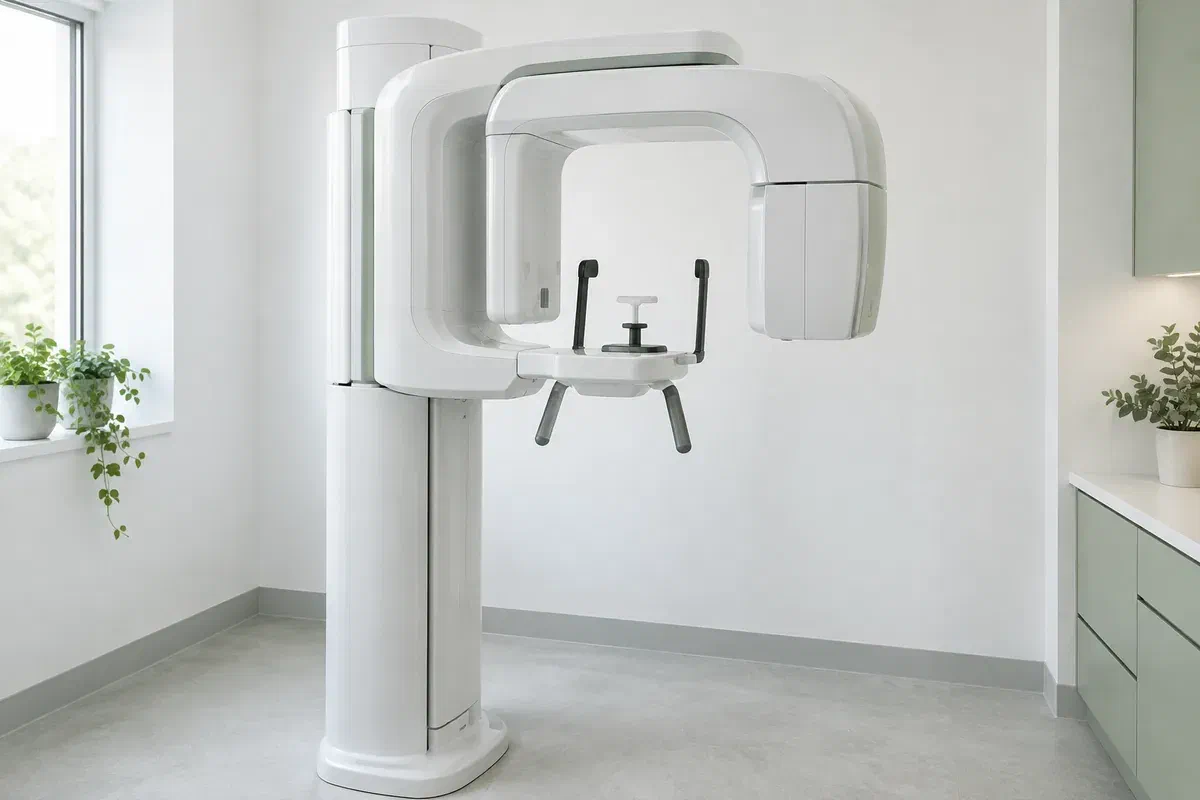
Illustrative discussion near generic panoramic imaging equipment, not actual clinic facilities
General & Family Dentistry

Panoramic Dental X-rays in Halton Hills

An ache near the back of the jaw can be hard to pin down: is it a tooth, the bone or something else? For people across Halton Hills, learn how a panoramic X-ray may help investigate, why describing the symptom matters more than naming a test, and what the image can’t tell you.

Halton Hills · investigating a symptom

Tell us what you feel. We’ll work out what to look at.

Discomfort that spreads along the jaw, a swelling that comes and goes, or a bite that suddenly feels different can leave you unsure where the trouble starts. In some of those situations, a dentist may consider a panoramic X-ray. It is one wide, flat image that takes in the teeth and both jaws, which makes it unlike the close-up films that focus on two or three teeth.

The symptom comes first; the picture follows only if it would help. Your dentist should decide based on what you describe, what the examination shows and any useful records already on hand. A wide view is not something you need simply because you are new or because some years have gone by. Feel free to ask what it would clarify.

We can discuss whether this view would help with your assessment, and how the imaging would be organised. Mention previous X-rays, any possibility of pregnancy and anything that makes standing or keeping still a challenge. Describing your symptom in detail, including when it began and what makes it worse, is the most useful thing you can bring.

Describe your symptom to our team
Illustrative unbranded panoramic dental X-ray unit in an empty imaging room
Clues that may point to imaging

From an ache to an answer.

Panoramic images help with certain questions about where teeth are and how the jaws appear. Your account of the problem and the examination findings guide whether this particular view fits, or whether another test should come first.

A buried tooth that may be involved

Sometimes discomfort near the back of the mouth relates to a wisdom tooth or another tooth that has not fully come in. The overview can show where it sits. Ask if more detail will be needed later; the image does not decide on its own whether a tooth should be removed.

A problem affecting more than one area

When symptoms or planned treatment involve several teeth or both sides of the jaw, a broad record can be useful. It is part of the picture rather than all of it. Ask what it will help decide and what will still need assessing.

Swelling, injury or a change in function

A knock to the face, recurring swelling or a jaw that opens differently may prompt the dentist to look more broadly. Explain the symptom in your own words rather than requesting a particular image, and let the examination point to the right test.

This kind of X-ray lays the curved jaw out flat, so some structures overlap and proportions can shift. Fine detail may be missing in places. The image does not stand in for a clinical examination, close-up radiographs or other investigations when those are appropriate.

How the broad view helps the search

Narrowing down where the problem might be.

Because it covers both jaws and the overall layout of the teeth in one image, a panoramic view can help a dentist see a symptom in context. No sensor goes inside your mouth for it, which differs from several smaller X-ray types. For a detailed question about a single tooth, another view may still be needed.

A finding only helps once it is interpreted alongside your symptoms and examination. Ask what in the image relates to your discomfort, whether anything needs another look, and what the view simply cannot show. A clear-looking picture does not mean every possible cause has been ruled out.

Patient information: RadiologyInfo — panoramic dental X-ray

Context around one complaint

Seeing the painful area alongside the rest of the jaw may make the explanation easier to follow. Ask how the overview affects what happens next. A broader image is helpful only when the question needs breadth.

Records for a specialist, if needed

If you are referred, the image and relevant notes can be shared where appropriate. Ask how and when that will happen, and whether the specialist will accept it. They will still examine you themselves.

A defined reason for further testing

The overview may explain the symptom or show that another investigation is needed. Ask what is still unclear before agreeing to more imaging. The next test should address a particular gap, not follow by default.

If imaging goes ahead

How it usually works, in plain terms.

Each imaging provider uses its own equipment and routine. Check which location will take the image, and mention anything affecting your posture or comfort before the booking is final.

  1. Link the image to your symptom

    Your dentist reviews your history, the examination and earlier records before recommending the view. Ask what it will show about your complaint and whether existing images would do. Let them know about pregnancy or possible pregnancy.

  2. Remove what might block the image

    Earrings, necklaces, glasses, hair accessories and removable dental pieces may need to come off. The operator shows you how to stand or sit at the unit. If your jaw is sore, your neck is stiff or your balance is unsteady, say so.

  3. Keep still as the arm moves

    The unit turns around your head for a short time. You will be told how to hold your lips, tongue and head. If opening or biting is painful because of your symptom, mention it before the exposure so the setup can be reconsidered.

  4. Review the findings with your dentist

    A clinician must read and explain the image before it can guide a decision. Ask who will go over it and whether further tests or referral are likely. If the X-ray was taken elsewhere, confirm how it will reach your dentist.

While you wait for a next step

Keep an eye on the symptom, too.

A panoramic X-ray needs no recovery period or aftercare in most cases. If other treatment was carried out at the same visit, follow the separate instructions.

Understand what was found

Ask whether the result explains your symptom, raises a fresh question or shows nothing further is needed. Have medical or dental terms explained. Knowing how the result shapes the plan is what makes it worthwhile.

Follow through on recommendations

Keep any further assessment or referral appointment and ask how results will be shared. An unexpected finding should be explained before you decide how concerned to be. Your dentist should separate what is known from what needs more information.

Note the date and location of the image

Future clinicians can then judge whether it is still useful for their question. Existing images sometimes prevent duplication, though that depends on their quality and on what has changed since.

Possible ways to investigate

A wide image is one tool among several.

Different types of imaging show different things. Ask why one is recommended for your symptom and how its likely benefit compares with the exposure involved.

When the problem may span the jaw

A panoramic overview

Suited to certain questions about tooth position and jaw structures. It uses ionizing radiation, so it should be justified clinically. It does not cover every detail other dental images can show.

When the symptom points to one tooth

Targeted intraoral radiographs

A small, focused image may answer a question about a particular tooth or its surrounding bone more clearly. Ask why that detail matters. The wide image does not automatically make bitewings or periapical views unnecessary.

When the cause is still unclear

Further imaging when it is justified

Some symptoms may call for another method, including CBCT where indicated. It is a separate examination, not an automatic next step. The dentist should name the unanswered question and explain why more information is needed.

Symptoms and imaging

Questions about using X-rays to find a cause.

Imaging should follow your examination and the information your care needs. Tell the team about earlier images and any concerns before the X-ray is taken.

(905) 876-4488
Can a panoramic X-ray show the jaw joints?
It may show part of the jaw joint area, but it has limits for assessing those joints in detail. If joint problems are suspected, the dentist will explain whether this view, another test or a referral is more appropriate.
My ache might be sinus-related. Would a panoramic show that?
The upper jaw sits close to the sinuses, and part of that region can appear on a panoramic image. It is not a full sinus examination, though. If a sinus cause is suspected, your dentist may suggest seeing your family doctor.
Should I take pain medication before the appointment?
Follow your usual advice from a pharmacist or doctor about over-the-counter pain relief. Tell the dentist what you have taken and when, as it can affect how symptoms present during the examination.
What if opening or biting hurts too much for the X-ray?
Tell the operator before the image is taken. They may be able to adjust the position, or the dentist may suggest a different approach. You should not be expected to hold a position that causes significant pain.
Could the X-ray miss a small cavity causing my pain?
Yes. The wide view has limited detail for some surfaces, especially between teeth. A clinical examination and close-up X-rays may be needed to find that kind of decay.
Do digital panoramic images use any radiation?
Yes. Digital units still use ionizing radiation, usually a small dose. The image should be clinically justified. ‘Digital’ describes how the image is captured and stored, not a lack of radiation.
Panoramic X-rays for Halton Hills

Start with what you are feeling.

Get in touch with our Bronte Street practice and describe your symptom, how long it has lasted and what helps or worsens it. Let us know about recent X-rays and any positioning needs. We will explain whether imaging would help and how the result guides your care.

We recommend imaging only where there is a clinical need. Pictures of equipment on this page are examples, not a confirmation of an on-site unit or a set appointment format.

Visit Milton Dental Clinic
Unit 1, 342 Bronte St. S, Milton, ON L9T 5B7
Get directions Clinic hours
Monday–Thursday
11am–7pm
Friday–Sunday
By appointment only

Areas we serve

  • Milton
  • Oakville
  • Mississauga
  • Georgetown
  • Halton Hills
  • Acton
  • Campbellville
  • Brampton
  • Burlington
  • Puslinch
  • Rockwood
  • Erin
  • Caledon
  • Etobicoke
  • Old Milton
  • Bronte Meadows
  • Timberlea
  • Dorset Park
  • Dempsey
  • Clarke
  • Beaty
  • Coates
  • Harrison
  • Scott
  • Willmott
  • Ford
  • Bowes
  • Cobden
  • Walker
  • Mountain View
  • Milton Heights
  • Hawthorne Village
  • Valley View
  • Fallingbrook
  • Nassagaweya
  • Trafalgar
  • Esquesing
  • Nelson
  • Brookville
  • Moffat
  • Glen Williams
  • Norval
  • Limehouse
  • Stewarttown
  • Ashgrove
  • Hornby
  • Silver Creek
  • Speyside
  • Scotch Block
  • North Oakville
  • River Oaks
  • Glen Abbey
  • West Oak Trails
  • Palermo
  • Bronte Creek
  • College Park
  • Joshua Creek
  • Uptown Core
  • Preserve
  • Iroquois Ridge
  • Lisgar
  • Meadowvale
  • Meadowvale Village
  • Churchill Meadows
  • Erin Mills
  • Central Erin Mills
  • Streetsville
  • East Credit
  • Creditview
  • Heartland
  • Sheridan
  • Mount Pleasant
  • Credit Valley
  • Huttonville
  • Churchville
  • Springbrook
  • Northwest Brampton
  • Bram West
  • Fletcher's Meadow
  • Fletcher's Creek South
  • Alton Village
  • Orchard
  • Millcroft
  • Headon Forest
  • Tansley
  • Brant Hills
  • Tyandaga
  • Kilbride
  • Lowville
  • Aberfoyle
  • Morriston
  • Arkell
  • Hillsburgh
  • Freelton
  • Carlisle
  • Waterdown
  • Flamborough
  • Mountsberg

How can we help?

Milton Dental Clinic

Have a question about your smile? Choose the option that works best for you.

For urgent dental concerns, please call.
Messages are reviewed during clinic hours.