

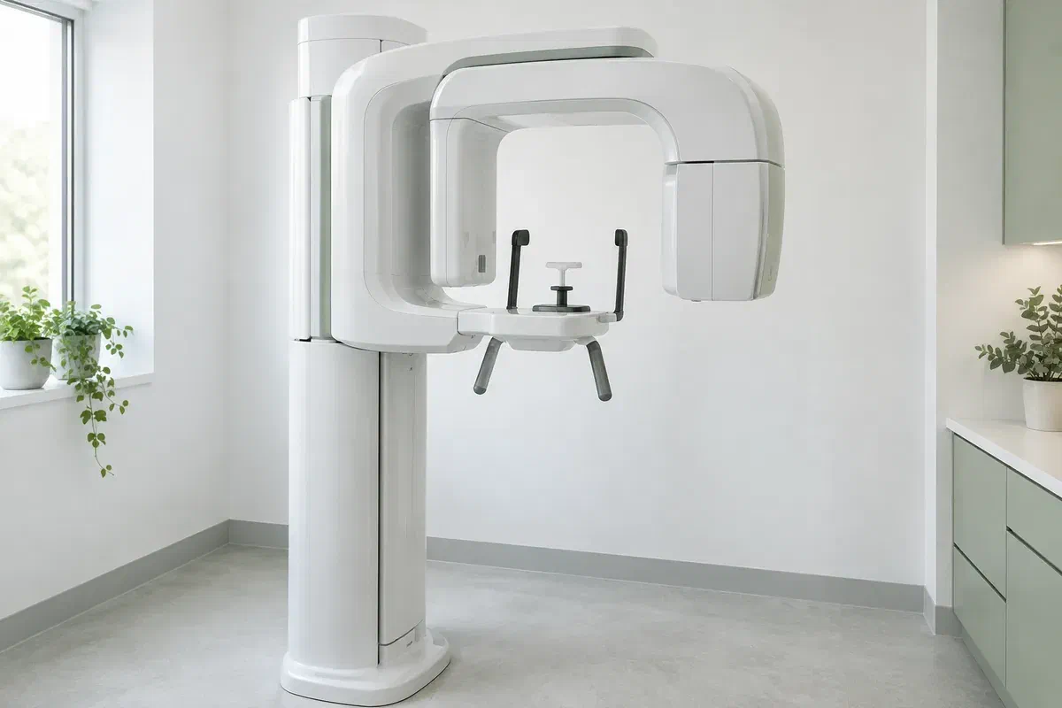
Illustrative discussion near generic panoramic imaging equipment, not actual clinic facilities
General & Family Dentistry

Panoramic Dental X-rays in Oakville

The machine that circles your head can feel unfamiliar the first time. Coming from Oakville, learn what a panoramic dental X-ray is for, what you will be asked to do while it runs, and how the image feeds into your dental plan.

For first-timers from Oakville

The X-ray that moves around you.

You stand or sit still, rest your chin on a small ledge, and an arm sweeps slowly from one side of your head to the other. That is a panoramic dental X-ray. The result is a single two-dimensional image showing the teeth together with the upper and lower jaws, which is quite different from the small films used to inspect a couple of teeth.

Knowing how it works is only half of it; knowing why it was suggested matters more. The dentist should tie the recommendation to something specific: findings from your examination, a symptom you raised, or a gap in the records already available. Time passing, or being new to the practice, does not by itself justify a wide image.

We can go over whether this kind of view suits your assessment and how the imaging would be organised. Mention any X-rays you have had recently elsewhere, any possibility of pregnancy, and anything that makes holding a position difficult. Sorting these out ahead of time keeps the visit simple.

Ask us what a panoramic X-ray would show
Illustrative unbranded panoramic dental X-ray unit in an empty imaging room
Why it might be suggested

Usually there is one of three reasons behind it.

A wide image is suited to particular questions about tooth position and the jaws as a whole. Your symptoms and the examination decide whether it is the right investigation, or whether a different approach would serve you better.

A tooth hiding below the gum

Wisdom teeth and other unerupted teeth can be difficult to assess by looking alone. An overview may show where they sit and how they relate to nearby teeth. Ask whether finer detail would be needed later; the image does not decide by itself that a tooth should come out.

Planning that reaches across the mouth

Treatment involving several teeth, or different areas of the jaws, can benefit from one broad record. It adds to the discussion rather than covering everything. Ask which part of the plan it informs and what still needs examining.

A symptom that needs a look

Pain, swelling, a fall or bump to the face, or a change in how your bite or jaw works can prompt an imaging question. Tell the dentist what you are experiencing rather than naming the X-ray you think you need, so the test can fit the problem.

Because a panoramic image lays a curved jaw out flat, parts of it overlap and some shapes are slightly distorted. It does not capture every small detail and is not a replacement for a clinical exam, targeted X-rays or other tests when they are warranted.

What the sweep captures

Breadth is the strength; detail is the trade-off.

The finished image shows both jaws and the layout of the teeth on one record. You will not hold a sensor in your mouth for it, unlike with some smaller types of dental X-ray. That breadth suits certain questions well, while close detail of a single tooth may still need another view.

A picture is only as helpful as the explanation that comes with it. Ask what part of the image relates to your concern, whether anything calls for a further look, and what the image is unable to show. Seeing your whole mouth on screen is not the same as having every dental condition checked.

Patient information: RadiologyInfo — panoramic dental X-ray

Orientation for a specific question

Viewing a tooth or finding in relation to the rest of the jaw can make treatment choices easier to understand. Ask how the overview applies to your case. More coverage is helpful when the question calls for it, not as a rule.

A shareable record for a referral

When another professional is involved, the image and relevant notes can be passed along where appropriate. Ask how it will be sent and whether a recent image elsewhere could be used. Specialists will still carry out their own assessment.

A reason for whatever comes next

The wide view may answer the question outright or reveal why another test is needed. Ask what is still unclear before you agree to more imaging. Any next image should address a defined limitation, not follow by habit.

Step by step

How a panoramic X-ray appointment usually goes.

Equipment and routines differ from one imaging provider to another. Ask where the X-ray will be taken, and raise anything that could affect your positioning before the booking is confirmed.

  1. Confirm the purpose

    The dentist considers your history, examination findings and any useful earlier images before recommending this view. Ask what it will add and whether something already on file would do. If you are or may be pregnant, say so, so the need and precautions can be discussed.

  2. Get ready at the unit

    You may be asked to take off earrings, necklaces, glasses or removable dental appliances. The operator shows you where to place your chin, hands and front teeth. Mention neck discomfort, dizziness, limited mobility or trouble following instructions so the setup can be adjusted.

  3. Keep still during the sweep

    The arm travels around your head for a short time while the image is captured. The operator tells you how to position your lips and tongue and reminds you to stay still. If something feels wrong, speak up before the exposure starts.

  4. Review the image with your dentist

    The picture still has to be read and explained before your assessment is complete. Ask who will walk you through it and whether a referral or further test may follow. If another provider took it, confirm how it will be sent to your dentist.

Afterwards

No recovery needed, but plenty to ask.

A panoramic X-ray on its own generally needs no aftercare or recovery time. Separate instructions apply if you had other treatment during the same visit.

Understand what the result means

Ask whether the image supports the proposed plan, raises a fresh question or shows that nothing more is needed for this concern. If a term is unfamiliar, ask for it in plain words. The result is most useful when you see how it affects your next choice.

Book the follow-up that is advised

Arrange any further assessment or referral that is recommended, and check how you will be told about results. An incidental finding needs an explanation; it should not be brushed aside or assumed to be serious. Ask what is known and what still needs clarifying.

Keep a note of the X-ray

Tell any future dentist when and where the image was taken so they can judge whether it remains useful. A previous record can sometimes avoid a repeat, depending on its quality, the new question and how much has changed since.

Other ways to look

Three kinds of imaging, three kinds of answer.

Each method reveals something different. Ask why a particular view has been recommended and how its expected benefit compares with the exposure involved.

Broad, two-dimensional coverage

A panoramic overview

Appropriate for some questions about the arrangement of the teeth and the structure of the jaws. It uses ionizing radiation and should serve a clinical purpose. It is not a substitute for every other type of dental image.

Finer detail in a small area

Targeted intraoral X-rays

A close-up image can sometimes answer a question about particular teeth or their supporting bone more clearly. Ask the dentist why that detail matters. Having a panoramic does not automatically rule out bitewings or other focused views.

For a question still unanswered

Further imaging if justified

Some situations call for a different method, such as three-dimensional CBCT where it is indicated. That is a separate examination rather than a routine upgrade. The recommendation should identify the remaining question and why more information is needed.

Common questions

What first-timers often want to know.

Imaging decisions should follow your examination and what your care requires. Tell the team about earlier X-rays and ask anything on your mind before the image is taken.

(905) 876-4488
How long does the machine actually take to go around?
The rotation itself is short, usually well under a minute, though positioning beforehand takes a little longer. The operator will tell you when it is about to start and when you can relax. Ask if you would like a practice run of the position first.
I feel closed in during scans. Is a panoramic X-ray enclosed?
No. The unit is open, and you stand or sit with the arm moving around the outside of your head. You can say if you feel uneasy before it starts. Let the team know about anxiety in advance so they can explain each step calmly.
Will I need to hold anything in my mouth that could make me gag?
The detector sits outside your mouth. Many machines use a small bite piece held between the front teeth to line things up, but nothing is pushed toward the back of the mouth. Tell the operator if even that feels difficult.
Does going to a new dentist mean a panoramic X-ray is automatic?
No. The choice of imaging depends on your history, what the examination shows, the question at hand and any useful existing records. Being a new patient is not a reason in itself. Ask what information the image would add.
Is a digital panoramic radiation-free?
No. Digital imaging still uses ionizing radiation, generally at a small dose. It should be justified and carried out appropriately. “Digital” describes how the image is recorded, not whether radiation is involved.
Is it worth combining the X-ray with a check-up to save a trip from Oakville?
It may be, if the imaging is clinically appropriate and the arrangements allow it. Ask when you book whether the X-ray, examination and results discussion can be grouped, and confirm where the image will be taken.
Panoramic dental X-rays for Oakville

Ask first. The X-ray can wait for a clear reason.

Call or book with our Bronte Street practice to discuss your concern and any imaging that has been mentioned. Let us know about recent X-rays and any positioning needs. We will explain the clinical purpose, confirm what can be arranged and show how the result will guide your care.

Imaging is recommended only when clinically needed. Equipment photographs are illustrative and do not confirm an on-site unit or any specific appointment arrangement.

Visit Milton Dental Clinic
Unit 1, 342 Bronte St. S, Milton, ON L9T 5B7
Get directions Clinic hours
Monday–Thursday
11am–7pm
Friday–Sunday
By appointment only

Areas we serve

  • Milton
  • Oakville
  • Mississauga
  • Georgetown
  • Halton Hills
  • Acton
  • Campbellville
  • Brampton
  • Burlington
  • Puslinch
  • Rockwood
  • Erin
  • Caledon
  • Etobicoke
  • Old Milton
  • Bronte Meadows
  • Timberlea
  • Dorset Park
  • Dempsey
  • Clarke
  • Beaty
  • Coates
  • Harrison
  • Scott
  • Willmott
  • Ford
  • Bowes
  • Cobden
  • Walker
  • Mountain View
  • Milton Heights
  • Hawthorne Village
  • Valley View
  • Fallingbrook
  • Nassagaweya
  • Trafalgar
  • Esquesing
  • Nelson
  • Brookville
  • Moffat
  • Glen Williams
  • Norval
  • Limehouse
  • Stewarttown
  • Ashgrove
  • Hornby
  • Silver Creek
  • Speyside
  • Scotch Block
  • North Oakville
  • River Oaks
  • Glen Abbey
  • West Oak Trails
  • Palermo
  • Bronte Creek
  • College Park
  • Joshua Creek
  • Uptown Core
  • Preserve
  • Iroquois Ridge
  • Lisgar
  • Meadowvale
  • Meadowvale Village
  • Churchill Meadows
  • Erin Mills
  • Central Erin Mills
  • Streetsville
  • East Credit
  • Creditview
  • Heartland
  • Sheridan
  • Mount Pleasant
  • Credit Valley
  • Huttonville
  • Churchville
  • Springbrook
  • Northwest Brampton
  • Bram West
  • Fletcher's Meadow
  • Fletcher's Creek South
  • Alton Village
  • Orchard
  • Millcroft
  • Headon Forest
  • Tansley
  • Brant Hills
  • Tyandaga
  • Kilbride
  • Lowville
  • Aberfoyle
  • Morriston
  • Arkell
  • Hillsburgh
  • Freelton
  • Carlisle
  • Waterdown
  • Flamborough
  • Mountsberg

How can we help?

Milton Dental Clinic

Have a question about your smile? Choose the option that works best for you.

For urgent dental concerns, please call.
Messages are reviewed during clinic hours.