
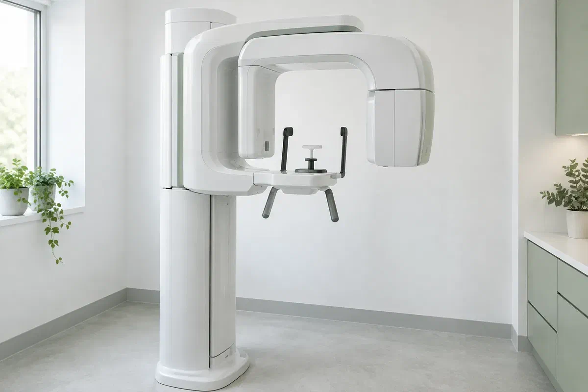
Illustrative discussion near generic panoramic imaging equipment, not actual clinic facilities
General & Family Dentistry

Panoramic Dental X-rays in Old Milton

Your last wide dental X-ray may be years old, filed at an office that has since changed hands. Find out how panoramic imaging is weighed in Old Milton, whether an older record still helps, and when a fresh view is actually needed.

Old Milton · panoramic imaging

Does the X-ray from years ago still tell the story?

Someone who has lived near Milton’s historic downtown for decades may have had a panoramic X-ray long ago and wonder whether it is still on file somewhere. That kind of image is a single flat picture that sweeps across both jaws and every tooth in them. Small, close-up X-rays do a different job, zooming in on just a few teeth.

Whether a new one is worthwhile depends on the question at hand. A dentist looks at your symptoms, what the examination turns up and which earlier records are available and still usable. Joining a new practice, or reaching a certain number of years since the last one, is not on its own a reason to repeat it.

We are happy to go over whether a wide view would help with your assessment and how the imaging would be arranged. Bring along what you know about past X-rays and where they were taken. Let us know about pregnancy or possible pregnancy, and about stiffness, balance or anything else that makes staying still harder.

Talk through your imaging history with us
Illustrative unbranded panoramic dental X-ray unit in an empty imaging room
Situations that call for the overview

Three questions a panoramic picture can help with.

This view is helpful for certain questions about the position of teeth and the overall shape of the jaws. What brought you in, and what the dentist sees during the exam, determine whether it fits or whether another approach is wiser.

A tooth that never fully came in

Some adults carry an impacted wisdom tooth or another buried tooth for years without trouble. A wide image may show where it lies now. Ask what it can confirm and whether more detail is needed, since the picture alone does not decide on removal.

Work planned across a larger area

Replacing several teeth or reviewing older restorations in different parts of the mouth can benefit from one broad record. It supports the discussion but is rarely the only thing needed. Ask what it contributes and what else must still be checked.

A change you can feel

Tenderness, swelling, a knock to the face or a jaw that moves differently may raise a question the dentist wants to explore. Describe the change rather than asking for a particular X-ray. Let the examination guide the choice of test.

The panoramic method projects curved anatomy onto a flat image, so structures overlap and some areas look stretched or narrowed. It misses fine detail in places and cannot take the place of an examination, targeted X-rays or other tests when those are called for.

What a broad record adds

A map of the whole mouth, read with care.

On one image, the dentist can see both jaws and how the teeth are spread across them. The sensor stays outside your mouth throughout, which sets it apart from bitewings and other small films. That reach is handy for particular questions, though a smaller view may still be better for detail.

Interpretation is where the picture becomes meaningful. Ask what in the image relates to your concern, whether anything deserves closer review, and what it cannot show. A wide picture should lead to a clearer explanation, not a belief that every dental issue has now been checked off.

Patient information: RadiologyInfo — panoramic dental X-ray

Perspective for a focused question

A tooth or a finding can be understood better when it is seen in relation to the whole jaw. That perspective helps when weighing treatment. Ask why the overview matters for you, rather than assuming more coverage is always an improvement.

Records that can travel

If another clinician joins your care, the image and relevant notes may be sent along where appropriate. Ask how that would work and whether an existing image is good enough. The receiving professional still examines you independently.

Grounds for any further step

The overview may settle the question or show why something more is needed. Ask what remains unclear before you agree to another image. Any extra test should target a specific gap, not follow simply because the first one was taken.

What to expect on the day

Four steps, start to finish.

Each imaging provider has its own equipment and routine. Ask where the X-ray will be done, and mention anything that might affect your positioning before the appointment is booked.

  1. Check whether an older image is enough

    Your dentist reviews your history, the examination and any earlier records before recommending a new view. Ask what a fresh image would add that the old one cannot. Mention pregnancy or the chance of it so the need and any precautions can be weighed.

  2. Remove items that show up on X-ray

    Jewellery, glasses, hair clips, hearing aids and removable dentures or partials often need to come out or off. The operator explains the supports on the machine. Speak up about neck pain, unsteady footing or limited movement so the position can be adapted.

  3. Stay steady as the unit rotates

    The arm travels around your head to collect the image. You will be guided on how to place your lips and tongue and how to stay still. If you cannot manage the position, say so before the exposure so the plan can change.

  4. Go over the findings together

    The image needs a clinician’s review and an explanation for you. Ask who will discuss it and whether a referral or further test is likely. If it was taken at a separate imaging location, check how it will get to your dentist.

After your X-ray

What should happen with the result?

There is normally no recovery period or special care after a panoramic image. If you had other treatment at the same visit, those instructions still apply.

Ask how the plan is affected

Find out whether the result supports what was proposed, opens a new question, or means nothing more is needed for this concern. Ask for unfamiliar terms in everyday language. The record matters most when you can see how it shapes the next decision.

Follow through on next steps

Keep any recommended appointments or referrals, and confirm how results will reach you. An unexpected finding deserves an explanation before you decide whether it is minor or concerning. Your dentist should be clear about what is certain and what is not.

Keep track of your imaging

Tell future dentists the date and location of the X-ray so they can consider whether it is still useful. Older images sometimes spare you a repeat, but that depends on their quality, the question being asked and how your mouth has changed.

Matching the image to the need

Not every question needs the wide view.

Each imaging method provides its own kind of information. Ask why one is being recommended and how its likely benefit compares with the exposure.

When the whole arrangement matters

A panoramic overview

Suitable for certain questions about where teeth are and how the jaws look. It uses ionizing radiation and should be taken for a clinical reason. It does not stand in for every other kind of dental X-ray.

When one area needs sharper detail

Focused intraoral X-rays

A small image may answer a question about a particular tooth or its supporting bone better than a wide one. The dentist should explain why that detail is important. Bitewings and other targeted views may still be needed after a panoramic.

When a question is still open

Other imaging only with a reason

Occasionally a different method, such as CBCT, is appropriate. It is its own examination with separate indications, not an automatic upgrade. A recommendation should state what remains unanswered and why the extra information is needed.

Old Milton imaging questions

Things people ask about the wide X-ray.

Decisions about imaging should rest on your examination and the information your care needs. Mention past images and ask your questions before the X-ray starts.

(905) 876-4488
Can my previous dentist send over an older panoramic image?
Often they can, with your permission. Your new dentist will look at its date, quality and whether it fits the current question. A usable record may reduce the need for a repeat, but sometimes a newer image is needed to see recent changes.
Do I take out my partial denture before the X-ray?
Usually, yes. Removable appliances with metal parts can block parts of the image. The operator will tell you what to remove. Bring a case to keep it safe while the picture is taken.
I wear hearing aids. Should I mention them beforehand?
Yes. Hearing aids may need to come out during the image, which can make spoken instructions harder to follow. Let the team know in advance so they can agree on simple cues, such as a tap on the shoulder or a hand signal.
Will a panoramic X-ray find all my cavities?
No. It is less detailed for fine areas such as the surfaces between teeth. An examination and close-up X-rays may still be needed to look for decay. Ask what the wide image is expected to cover for you.
Is a panoramic X-ray just a flatter version of a 3D scan?
It is a different examination. Panoramic imaging produces a two-dimensional picture; CBCT gives three-dimensional information and is used for its own reasons. The dentist should choose based on the question, not on the idea that 3D is always superior.
What if my balance makes standing at the machine difficult?
Tell the clinic or imaging provider before the appointment. Some units can be used seated, but setups differ, so ask what is possible. You should not be asked to hold a position that feels unsafe or painful; another plan may be better.
Wide-view X-rays near Old Milton

Bring your questions, and your X-ray history.

Reach out to our Bronte Street practice, close to Milton’s historic downtown, to talk about your concern and any suggested imaging. Share what you know about past X-rays and any positioning needs. We will explain the purpose, confirm arrangements and show how the result guides your care.

We recommend imaging based on clinical need. The equipment photos shown are illustrative and do not confirm an on-site unit or a particular appointment arrangement.

Visit Milton Dental Clinic
Unit 1, 342 Bronte St. S, Milton, ON L9T 5B7
Get directions Clinic hours
Monday–Thursday
11am–7pm
Friday–Sunday
By appointment only

Areas we serve

  • Milton
  • Oakville
  • Mississauga
  • Georgetown
  • Halton Hills
  • Acton
  • Campbellville
  • Brampton
  • Burlington
  • Puslinch
  • Rockwood
  • Erin
  • Caledon
  • Etobicoke
  • Old Milton
  • Bronte Meadows
  • Timberlea
  • Dorset Park
  • Dempsey
  • Clarke
  • Beaty
  • Coates
  • Harrison
  • Scott
  • Willmott
  • Ford
  • Bowes
  • Cobden
  • Walker
  • Mountain View
  • Milton Heights
  • Hawthorne Village
  • Valley View
  • Fallingbrook
  • Nassagaweya
  • Trafalgar
  • Esquesing
  • Nelson
  • Brookville
  • Moffat
  • Glen Williams
  • Norval
  • Limehouse
  • Stewarttown
  • Ashgrove
  • Hornby
  • Silver Creek
  • Speyside
  • Scotch Block
  • North Oakville
  • River Oaks
  • Glen Abbey
  • West Oak Trails
  • Palermo
  • Bronte Creek
  • College Park
  • Joshua Creek
  • Uptown Core
  • Preserve
  • Iroquois Ridge
  • Lisgar
  • Meadowvale
  • Meadowvale Village
  • Churchill Meadows
  • Erin Mills
  • Central Erin Mills
  • Streetsville
  • East Credit
  • Creditview
  • Heartland
  • Sheridan
  • Mount Pleasant
  • Credit Valley
  • Huttonville
  • Churchville
  • Springbrook
  • Northwest Brampton
  • Bram West
  • Fletcher's Meadow
  • Fletcher's Creek South
  • Alton Village
  • Orchard
  • Millcroft
  • Headon Forest
  • Tansley
  • Brant Hills
  • Tyandaga
  • Kilbride
  • Lowville
  • Aberfoyle
  • Morriston
  • Arkell
  • Hillsburgh
  • Freelton
  • Carlisle
  • Waterdown
  • Flamborough
  • Mountsberg

How can we help?

Milton Dental Clinic

Have a question about your smile? Choose the option that works best for you.

For urgent dental concerns, please call.
Messages are reviewed during clinic hours.