A ridge too slim or short
On a scan, the bone may not have enough width or height for an implant in the position the crown needs. Nearby anatomy can limit the choice further. Seeing the cross-section often makes the recommendation easier to grasp.


The gum over a missing tooth can look perfectly healthy and still sit on thin bone. From Georgetown, find out how scans and examination guide decisions about ridge augmentation, and what the results might mean for your replacement.
Run your tongue over a healed gap and it may feel smooth and full. Underneath, the ridge of bone that once held the tooth could be narrower than it seems. That bone is what an implant or other replacement relies on, and augmentation is a way to build it up when it falls short of the plan.
This is why imaging so often comes before any grafting advice. A scan or X-ray can show the width, height and angle of the ridge and its relationship to nerves, the sinus and nearby roots. The findings, not the look of the gum, decide whether a graft is worth discussing.
When you come to our Bronte Street practice from Georgetown, we start with the replacement you are hoping for and examine the site. We will explain which imaging would help, whether a specialist should review it, and how any ridge treatment would link to the final tooth.
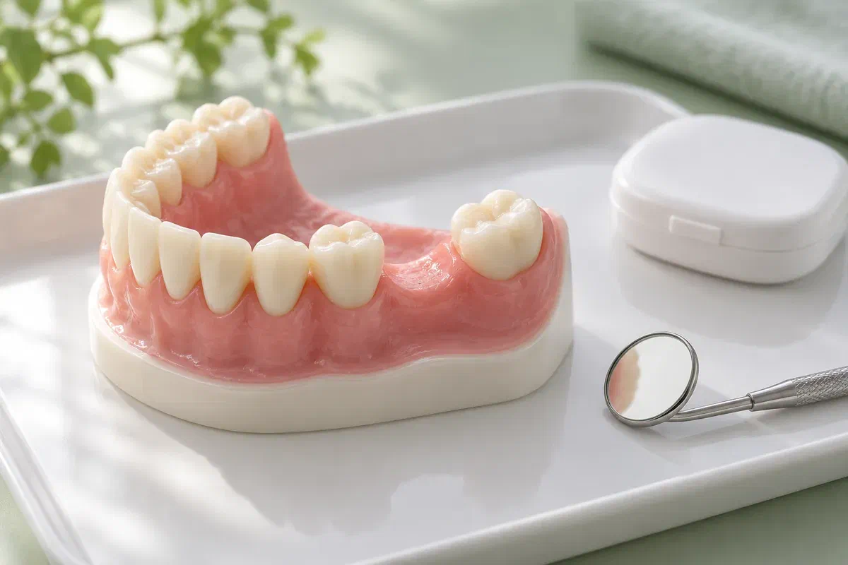
Examination and imaging together show the ridge’s shape, the structures around it and how the planned tooth would sit. Ask your clinician to point out the finding that matters most.
On a scan, the bone may not have enough width or height for an implant in the position the crown needs. Nearby anatomy can limit the choice further. Seeing the cross-section often makes the recommendation easier to grasp.
Some concerns show up more in the mirror than on a scan, such as a hollow beside a bridge or partial. Describe what you notice. Bone, gum thickness and the design of the restoration may each play a part.
Images can show the effects of past extractions, grafts, implants or injury. Bring any older X-rays you have for comparison. History explains a lot, but the current images and examination decide what happens next.
A thin-looking ridge on an image does not always mean grafting. The need depends on the specific replacement planned.
If augmentation is indicated, it aims to give the planned treatment a site that suits it, such as enough bone to place an implant where it should go or a ridge outline that supports a natural-looking tooth. Ask what the images suggest would change after grafting.
Grafting comes with surgery, a healing period, review visits and, at times, further treatment. Follow-up imaging may also be needed. The amount of new bone cannot be guaranteed, and a successful graft does not ensure a successful implant. A sound plan sets this out clearly.
Patient information: AAP — ridge modification for implant planning
Ask the clinician to show where the future tooth would sit on your images and where the bone falls short. That comparison makes the reason for grafting concrete.
Some grafts are placed at the same time as an implant, and others heal first. Ask which approach your findings support. The interval affects planning for work and a temporary tooth.
Materials and membranes differ between procedures. Raise any preferences about their origin early on, and ask whether a different choice would affect the plan or the result.
The approach and timing vary with what the imaging shows and the restoration planned. Ask who interprets your images and who carries out each stage.
The gap is examined and the right imaging arranged, alongside a review of your gums and medical history. Mention smoking, medications, earlier grafts, or trouble healing in the past. Share what you want from the new tooth.
The clinician walks you through the images, the proposed technique, materials, recovery, fees and alternatives. Ask about donor sites, membranes, and any later removal visit. Agree how the gap will be managed meanwhile.
If you proceed, the area is treated with the comfort plan and method discussed. Written instructions for your procedure follow. Recovery varies, so rely on your own guidance rather than a friend’s experience.
After healing, the site is checked and further imaging may be taken before the next stage. A comfortable gum is not enough evidence on its own. Ask what the clinician expects to see before moving on.
Keep your aftercare sheet and contact numbers close. Ask whether exercise, cleaning or an appliance could place pressure on the treated area.
A partial or denture may need adjustment or a pause in wear. Do not assume it can rest on the area as before. Settle how you will manage meals and appearance before treatment.
You will be given cleaning steps for the site and your other teeth, plus guidance on food. Avoid pulling your lip down to look or pressing on the area. Check before you change the routine.
Go to reviews even when you feel well. Contact the treating team about pain that worsens, bleeding that returns, discharge, or an opening in the wound, so they can decide whether to see you.
Patient information: Cambridge University Hospitals — bone grafting for implants
We can show you your images and explain them with a model. Ask about recovery and the final tooth as well.
An assessment at our Bronte Street practice gives you a proper look beneath the gum. We will examine the gap, explain which imaging would answer your questions, and relate the findings to the tooth you want, including whether grafting or a different plan makes more sense.
If a site that has already been treated changes, use your treating clinician’s contact instructions or call us for advice.
