
Illustrative patient discussing an oral lesion assessment with a clinician
Specialized Care

Oral Lesion Removal

A lump, patch, or persistent sore in the mouth should be assessed before deciding on removal. Our Milton team can explain the findings, discuss whether tissue sampling or treatment is appropriate, and help clarify any specialist referral and follow-up needed.

A closer look at oral lesion removal

Understand the tissue change before deciding what should be removed.

Oral lesion is a broad term for an area of tissue that appears different from its surroundings. It might be noticed as a lump, a patch, a sore, or a change in texture. Those descriptions do not establish a diagnosis, and they do not mean that every area should be removed immediately or treated in the same way.

Removal may be appropriate for a selected lesion, but the first step is an examination and a reasoned plan. Sometimes a sample is needed before further treatment is chosen. In other circumstances, a small area may be removed as an excisional biopsy, or a specialist may need to assess the location and findings first.

At Milton Dental Clinic, tell us what you have noticed and what concerns you most, whether that is repeated irritation, uncertainty about the diagnosis, or a previous recommendation. We can discuss the assessment and referral pathway without assuming that a consultation guarantees same-day removal or a particular result.

Discuss a mouth tissue change
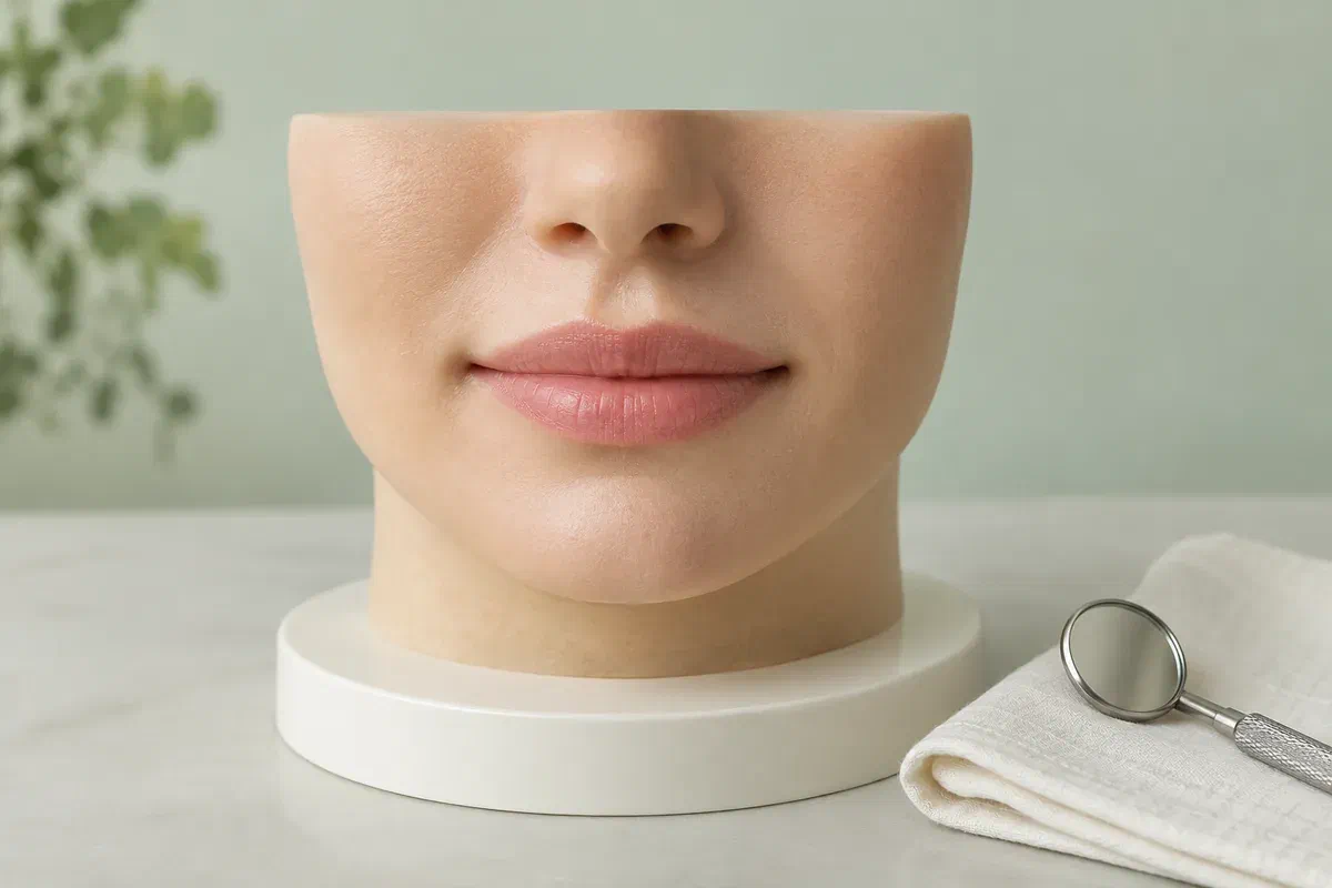
Illustrative closed-mouth teaching model on a display base beside a dental mirror
Information to bring to the examination

The history of the area helps explain what needs investigating.

Describe when the change began and whether it has grown, returned, or behaved differently. If another provider has already assessed it, bring the report or referral so that important details do not have to be reconstructed from memory.

An area that is repeatedly irritated

Explain whether the tissue catches between teeth or rubs against a restoration, denture, or another surface. The dentist can assess both the lesion and the possible source of irritation. Do not assume that an obvious rubbing point proves what the lesion is or makes investigation unnecessary.

A persistent or changing lump, patch, or sore

Mention changes in size, colour, texture, or sensation, and whether the area bleeds or affects eating. A painless lesion can still need assessment. Avoid repeatedly pressing, biting, or scraping it while trying to determine whether it will disappear.

A previous sample, removal, or recurrence

Tell the team if the area has been treated before and whether a laboratory result was issued. A returning lump should be reassessed rather than assumed to be identical to the original lesion. Earlier records can help the clinician decide what information is still needed.

Arrange assessment of a persistent or changing mouth lesion and follow the urgency of any existing referral. Do not cut, burn, drain, or tie off tissue at home. Severe uncontrolled bleeding or difficulty breathing or swallowing requires urgent medical care.

What a clear plan should provide

Know the purpose of treatment and the questions that remain afterward.

Before a procedure, ask whether the goal is to obtain a diagnosis, remove a confirmed problem, reduce repeated irritation, or address more than one concern. Understanding that purpose helps you judge the recommendation and its alternatives. It also avoids the assumption that removing what you can see automatically completes all necessary care.

The plan should account for the location as well as the lesion itself. Ask about the nearby structures, expected healing, and any effect on eating or speaking during recovery. If specialist care is recommended, clarify what makes that setting appropriate and who will coordinate the referral and results.

A decision based on examination rather than appearance alone

The clinician considers the history and physical findings before advising on removal. Ask whether further investigation could change the approach. Comparing the area with an internet photograph cannot establish that it is harmless or determine which procedure would be suitable.

An explanation of the tissue examination plan

Ask whether removed tissue will be sent for laboratory assessment and how the findings will be explained. If a biopsy has already been performed, discuss how its result affects the treatment plan. Make sure you understand which provider will take responsibility for receiving and reviewing the report.

Follow-up that includes both healing and diagnosis

A review may need to assess the wound, discuss laboratory information, or plan further care. Ask which of these purposes applies to each appointment. A comfortable healing site does not answer every diagnostic question, and a report may still need discussion after the area looks settled.

From assessment to an agreed procedure

Explain the findings, choose the setting, and plan the result discussion.

You should have an opportunity to understand the recommendation before consent. Tell the clinician if you are worried about the procedure, the diagnosis, or the effect on everyday activities so those concerns can be addressed directly.

  1. Review the lesion and relevant background

    Describe its timeline and any previous treatment, and bring available pathology reports or referral details. Review medicines, allergies, bleeding history, and medical conditions. The examination helps determine whether removal, sampling, observation, or specialist assessment is appropriate.

  2. Discuss the proposed extent and alternatives

    Ask what tissue would be removed and why that approach fits the findings. Review site-specific risks, the anaesthetic plan, and whether a specialist should perform the procedure. Confirm the likely stages of care and fees rather than assuming that the initial visit includes treatment.

  3. Carry out the agreed treatment and explain wound care

    If removal proceeds, the treating clinician manages the area according to the chosen procedure and sends tissue for examination when indicated. Ask whether stitches are used and how they will be managed. Before leaving, obtain clear instructions and a contact route for recovery concerns.

  4. Review healing, findings, and any further action

    Attend the planned follow-up and ask for the result in plain language. Clarify whether the original concern has been resolved or whether additional assessment, treatment, or monitoring is recommended. If a local source of irritation remains, discuss how it should be addressed.

After lesion removal

Protect the treated area and keep the next step in view.

The amount of tissue removed, its location, and your health influence recovery. Follow the written instructions supplied by the treating clinician and ask about any part that is difficult to manage at home.

Use the specific eating and cleaning advice

Ask how to clean near the site and which foods or activities should be avoided temporarily. Take care around numb tissues after local anaesthetic. Do not assume that instructions for a different dental operation apply to this wound or begin an unadvised rinsing routine.

Avoid disturbing the wound or stitches

Do not pull the lip or cheek repeatedly to inspect the area or pick at the healing surface. Clarify whether stitches dissolve or require removal. If something appears unusual, describe it to the team and follow their advice rather than attempting to trim or remove it yourself.

Report concerning changes and complete the review

Contact the provider about worsening pain, significant swelling, fever, or bleeding that does not settle with the instructions given. Keep the results arrangement even if recovery feels straightforward. If the expected contact does not occur, call to confirm how the findings will be discussed.

Choosing the next step

Removal is one route; the diagnostic question guides the decision.

Ask why the recommended pathway is appropriate for this particular area. The plan should explain what is being investigated and what would cause the clinician to recommend a different approach.

When the examination supports monitoring

Observation with a defined review

The clinician may recommend review after addressing a relevant source of irritation. Ask when the area should be reassessed and what changes require earlier contact. Monitoring should have a specific rationale and endpoint, rather than becoming indefinite delay of a recommended investigation.

When tissue examination or local treatment is indicated

Sampling or removal of an appropriate lesion

A partial sample or complete removal of a suitable small lesion may be discussed. Ask which approach is intended and whether more treatment could follow the result. The visible size alone does not determine the diagnosis or guarantee that one procedure will complete care.

When the findings or location require wider expertise

Specialist assessment before definitive treatment

A referral may be the appropriate first step when the lesion needs further evaluation or a different procedural setting. Clarify the referral's urgency and how to follow it up. A specialist consultation can help establish the right treatment sequence before tissue is altered.

Oral lesion removal questions

Clarify the purpose of removal and what must happen afterward.

The answers depend on the examination and any existing diagnostic information. Bring previous reports and explain your main concern so the consultation can focus on the decisions relevant to your lesion.

(905) 876-4488
Can every small mouth lump be removed at the first consultation?
No. The clinician first needs to assess the area and decide whether removal is appropriate in that setting. A sample, further records, or specialist opinion may be needed. Ask what the initial appointment includes and which steps would precede treatment.
Is removing an oral lesion always the same as diagnosing it?
No. Removal may provide tissue for laboratory examination, but the diagnostic process includes interpreting that result with the clinical findings. Ask how the report will be reviewed and whether further care could be recommended after the visible area is removed.
Should a denture that rubs the lesion also be assessed?
Yes. Explain the rubbing and bring the denture to the consultation. The clinician can assess both the tissue and the appliance. An apparent irritation source should not replace investigation of the lesion, but it may be relevant to the overall treatment plan.
Does complete removal mean an oral lesion can never return?
That cannot be guaranteed. The outlook depends on the diagnosis, procedure, and contributing circumstances. Ask what monitoring is recommended and report a returning or changing area. A recurrence should be assessed rather than treated at home or assumed to be harmless.
Will I need a results discussion if the removal site heals normally?
Yes, when tissue was sent for examination or a follow-up discussion was arranged. Healing does not reveal the laboratory diagnosis. Confirm who will contact you and call if the expected date passes without an explanation of the findings.
Can I use a home skin-tag remover on a lump inside my mouth?
No. Do not cut, burn, or tie off oral tissue or apply a product intended for skin lesions. The area needs a dental or appropriate medical assessment first. Home removal can injure tissue and interfere with establishing what the lesion is.
Oral lesion removal assessment in Milton

Let us understand the change and help clarify the appropriate pathway.

Call our Bronte Street practice at (905) 876-4488 to discuss an oral lesion assessment. Mention an existing referral, previous biopsy, or recent change when booking so the team can help plan the relevant next step.

Removal is not appropriate for every lesion. Diagnosis, treatment setting, referral, recovery, and follow-up depend on the examination and any laboratory findings.

Visit Milton Dental Clinic
Unit 1, 342 Bronte St. S, Milton, ON L9T 5B7
Get directions Clinic hours
Monday–Thursday
11am–7pm
Friday–Sunday
By appointment only

How can we help?

Milton Dental Clinic

Have a question about your smile? Choose the option that works best for you.

For urgent dental concerns, please call.
Messages are reviewed during clinic hours.